A Writer with a Day Job

Dengue_map.jpg

I’m going on a bit of a tangent this week and breaking away from my usual theme of forensics and writing to touch on another aspect of my life—my day job. Like many writers out there, writing may be my passion, but I have a responsibility to help my husband support our family financially, so I work full time. I worked in the field of HIV research for 20 years, but last winter my lab downsized, and I started looking for a new position. Last July, I joined a dynamic research group specializing in infectious diseases. We study a range of diseases, including pneumococcal infections and influenza, but our big project is a very large, international study of dengue fever.

In the map above, the areas in blue indicate the current risk for dengue virus infection, and the pins in red indicate areas where the disease is spreading or has been carried by travellers. Currently 40% of the world’s population lives in areas where the virus is endemic, leading to an estimated 50 million cases of dengue fever annually. Of those cases, 500,000 patients require hospitalization and 20,000 – 25,000 patients die of the disease. The virus spreads to humans by two types of mosquitos and the incidence of the disease matches the geography inhabited by these insects. Because of global warming and the northern spread of the Aedes Albopictus mosquito into the southeastern United States, the CDC has classified the dengue virus as a Biodefense Category A pathogen; a category that encompasses the most dangerous of the infectious diseases due to their easy transmission, high mortality rate and lack of effective treatment.

The large majority of patients infected with dengue virus show no symptoms at all or only present with a mild illness including fever, aches, a mild skin rash and joint pain—leading to the colloquial name for this disease: bone break fever. But approximately 5% progress to severe illness, and a subset of those exhibit life-threatening disease (dengue hemorrhagic fever and dengue shock syndrome), including symptoms such as platelet loss, abdominal bleeding, fluid accumulation in the chest, low blood pressure and organ dysfunction. There are four main serotypes of dengue virus, but previous infection with one serotype doesn’t protect you from the other three. In fact, a subsequent infection with a different serotype significantly increases the chances of life-threatening disease. Once infected, there is no effective anti-viral treatment, and all hospital staff can do is keep the patient hydrated.

An electron micrograph showing a cluster of dengue virus particles

An electron micrograph showing a cluster of dengue virus particles

The mystery with dengue infection is why there is so much variability in the range of symptoms. Over 80% of those infected have either no symptoms (many don’t even know they’ve ever been infected) or only very mild symptoms. So what causes some people to progress to severe or fatal disease? Our team hypothesizes that there are common genetic variations in certain genes that affect the immune system and how it responds to the infection, and it is these variations that predispose some individuals to dengue hemorrhagic fever. To this end, we are studying over 9,000 participants from 10 international sites including Mexico, Nicaragua, Vietnam, Columbia and Sri Lanka over a 5 year period. We’ll look at the patients’ DNA, RNA and serum to identify variations in their genes and antibodies. Ideally, we’ll find a specific gene or genes that affect the way the body reacts to dengue infection.

The long-term goal in dengue research has always been to produce a vaccine or treatment that will assist those most at risk for serious infection. Hopefully, armed with this information, we’ll be able to drastically reduce the number of 25,000 dead annually.

Photo credit: DengueMap and Wikimedia Commons

Forensic Case Files: The Princes in the Tower

Princes in the tower.jpg

In the last Forensic Case File, I discussed the remains of Richard III, finally discovered in Leicester (pending confirmation). Richard III is infamous for supposedly killing his two nephews, Edward V, 13, and his younger brother Richard, 10, the sons of Edward IV. But because Parliament ruled Edward IV’s marriage invalid and his sons illegitimate, the crown legally went to Richard III in June of 1483. That month, Richard sent his two nephews to the Tower of London. They were seen playing on the grounds over the following months, but were never seen again after the summer of 1483. Surely they had been killed, but the question was by who and when?

The two most famous accounts of the deaths of the young princes were written by Thomas More, Henry VIII’s Lord Chancellor, and William Shakespeare, the famous poet and playwright. Both accounts paint Richard III with a dark brush, portraying him as a villainous monster. But the truth of the matter is that both accounts were written by men who were never witness to the supposed crime—More was eight years old when Richard fell in battle, and Shakespeare was born seventy-nine years later. History tells that More’s account of Richard III came from John Morton, the Archbishop of Canterbury during Henry VII’s reign and a sworn enemy of Richard III. It’s a well-known fact that history is written by the victors; in this case, the House of Tudor was legendary for removing any and all rivals to the throne. It’s not much of a stretch to suppose that the story of Richard III as a menacing hunchback who murdered his nephews was used to blacken the name of a defeated rival family, the House of York.

The truth of the matter is that Richard III had no concrete reason to kill the boys and nothing to gain from their deaths. His claim to the throne came with the approval of both the English public and Parliament. Had Edward IV’s marriage been upheld and his royal line remained intact, there were three other male heirs closer to the throne than Richard, so simply killing his nephews would not have assured him a place as monarch.

So if Richard III wasn’t responsible, who else stood to gain from the princes’ deaths?

In her classic 1951 novel, The Daughter of Time, novelist Josephine Tey makes a convincing argument that Henry Tudor had to most to gain following Richard III’s death at the Battle of Bosworth Field when he was crowned Henry VII.

Henry never missed an opportunity to vilify Richard posthumously, yet when he came to London he never remarked on the fact that the boys were missing from the Tower. It’s been suggested that he would have happily pinned the disappearance and murder of the boys on the fallen king simply to make himself look more favourable by comparison. But perhaps he didn’t want to bring the princes’ disappearance to light because he had ordered their deaths. The boys were also a threat to him—following the death of Richard III, Henry VII repealed the Titulus Regius statute that made Richard III king, nullifying Richard’s reign while simultaneous declaring Edward V King of England and his brother the heir. The boys were a direct threat to his reign as monarch. The boys were never seen after 1483, but it is possible that they were still alive in the Tower as late as 1485 when Henry VII arrived in London. Killing the princes would have ended the last remaining hope that the House of York would be able to reclaim the throne.

Officially, the murder has been laid at the feet of James Tyrrell, an English knight in the service of the House of York and under Richard’s command. Nearly twenty years after the rise of Henry VII, Tyrrell supposedly confessed to smothering the boys and was hanged for his crime. But there was never any transcript of the confession, no formal charges were laid, nor was there ever a trial. In fact, the account was only released after Tyrrell himself was already dead. According to the reported confession, the only witness to the crime conveniently died at the Battle of Bosworth Field along with Richard III.

In 1674, nearly 200 years after the princes’ disappearance, the skeletons of two children were found in a wooden box beneath a stone staircase during renovations in the White Tower. Since it was assumed that these were the remains of the missing princes, the bones were interred with royal honour in an urn in one of the walls of the Henry VII Chapel in Westminster Abbey.

In 1933, Professor William Wright, one of the foremost anatomists of his day, was granted permission to examine the remains. Inside the urn Dr. Wright found the incomplete remains of two children he determined to be between the ages of 7–11 and 11–13 years of age, mixed with several animal bones and a handful of rusty nails. There was no sign of any skeletal trauma on the remains, supporting the theory that the princes had been suffocated. But as the sex could not be determined on the pre-pubescent bones and DNA analysis was decades away, that was the only information learned and the bones were re-interred. Current osteological research now gives scientists methods to determine age of pre-pubescent individuals (although it remains a somewhat inexact science) and DNA analysis could identify the remains if a female family member could be found. Since relatives found to test against Richard III’s remains are not in the same female line as Edward IV’s sons, a new family member in the maternal line would have to be located for confirmation.

So, the mystery surrounding the death and identity of the lost princes remains. If scientists are allowed to examine the remains again, some new answers may come to light. But, unfortunately, it remains a real possibility that the fate of the princes may remain lost to the ages.

Photo credit: Wikimedia Commons

Forensics 101: DNA Profiling for Identification

VNTR gel.gif

Last week, I covered DNA as a tool for identifying remains. This week, I’m going to discuss how scientists test DNA to prove an identifying match.

DNA strands contain different regions, many of which are genes that code for essential protein products. But a very large proportion of sequences don’t code for any currently known genes. These sequences also contain short tandem repeats (STR)—small snippets of DNA that are two to six base pairs long and repeat from three to one hundred times in a row. The locations of these repetitive sections are called variable number tandem repeats (VNTR). Genetically speaking, unrelated individuals will have different numbers of repeated STR segments at known VNTR locations, but related family members will share similar numbers of repeated segments. Since human offspring share a combination of traits from both parents, the power of DNA profiling lies in analyzing numerous segments to definitively prove identification. In North America, it’s standard protocol to analyze thirteen specific locations simultaneously.

Scientists use the polymerase chain reaction (PCR) to examine known VNTR locations. PCR is an assay used to amplify small amounts of specific DNA sequences so they can be visualized later on a gel (we’ll look at PCR in more detail in a future Forensics 101 post). The picture above illustrates typical PCR results. In this case, a gel shows the difference in length of the D1S80 VNTR location of six unrelated individuals, flanked on each side by a marker of known size. As you can see, the pattern for each individual subject is different in each vertical lane.

An example of DNA profiling between a father (1), mother (3) and child (2).

An example of DNA profiling between a father (1), mother (3) and child (2).

There are two types of matches in DNA profiling—identity and inheritance. In identity matching, the unknown sample is tested against a known individual’s DNA. If the two samples match exactly, then the unknown person is identified as the known donor. In inheritance matching, the unknown sample is tested against a sample drawn from a potential family member. If parents are used as donors, then each band in the unknown sample must match with one of the two parents. If a more distant relation is used, then degrees of relatedness are calculated into the expected results.

The bottom line of this testing is that the unknown sample must be tested against a sample (blood, hair, tissue etc.) of known origin. In the case of Richard III’s ancestors, separated by five centuries from the king himself, genomic DNA profiling as described above simply wouldn’t be feasible due to the number of generations separating Richard III and his current ancestors. But when this same method is applied to mitochondrial DNA, because of the consistency of maternal transmission, identical or near identical results are expected between family members, even those separated by multiple generations.

Next week, we’ll look at the historical details surrounding the Princes in the Tower, the young boys Richard III is accused of murdering. Nearly one hundred years after their disappearance, bones were recovered at the Tower of London. But where they of the missing princes? See you next week to find out…

Photo credit: PaleWhaleGale and Magnus Manske

Forensics 101: Tracing Lineage Through Maternal Mitochondrial DNA

DNA-molecule3.jpg

Deoxyribonucleic Acid (DNA)Last week in a post on the potential discovery of Richard III’s remains, I wrote that scientists hope to confirm the identity of the remains based on DNA, specifically mitochondrial DNA. Over the next few weeks, I’m going to explain DNA identification, starting with the basics and then branching out into how it is used to name the dead.

DNA (deoxyribonucleic acid) is the code of life, the genetic information contained in every cell in the form of twenty-three pairs of chromosomes (except for sperm and eggs cells which have twenty-three single chromosomes). The information contained in this genomic DNA specifies everything about us, from how we look to which health problems we’ll have an increased likelihood of developing. But there is also another kind of DNA contained in our cells—mitochondrial DNA (mtDNA).

Mitochondria are the power houses in our cells, the organelles or cellular structures that are responsible for producing the chemical energy required for every cellular activity—from protein production to ion transport to cellular reproduction. But unlike other organelles, mitochondria have their own unique tiny DNA genome—mtDNA. Due to the process of human fertilization, these small bits of DNA are transmitted in family lineages only through the maternal line.

Electron micrograph of two mitochondria

Electron micrograph of two mitochondria

An egg cell is a stripped down version of a typical cell in the body; essentially it only contains a nucleus carrying twenty-three single chromosomes plus several hundred mitochondria in the cytoplasm. A sperm cell is a small sack of DNA attached to a long tail for mobility, the base of which is packed with energy producing mitochondria to fuel the journey. At the moment of fertilization, the tiny sperm head fuses with the egg membrane and injects its DNA while the tail drops off and is lost. As a result, the only DNA inserted into the egg cell is the genomic DNA contained within the sperm head. All mitochondrial DNA that is then replicated as the fertilized zygote splits from one cell into approximately one hundred trillion cells comes solely from the mother. Fortunately, due to the nature of its sequences, mtDNA has a very low mutation rate. In other words, the same mtDNA is passed from grandmother to mother to child through the generations. Genetic testing of specific sequences of those mtDNA samples, even if separated by many generations, can definitively prove a family match.

Due to the number of copies of mitochondrial DNA in each cell (providing approximately five hundred identical copies of each gene versus two copies on the cell’s genomic DNA), forensic anthropologists often are more successful recovering mtDNA than genomic DNA from ancient and historical samples. So the combination of DNA yield and the consistency of the maternal line can provide identification, even for remains over five hundred years old.

Next week we’ll look more specifically at the kind of testing used to make a genetic match between samples providing conclusive DNA evidence for identification.

Photo credit: ynse and Louisa Howard

Forensic Case Files: Richard III's Remains Finally Found in Leicester?

Richard III, who ruled England from 1483 to 1485, was the last of the legendary Plantagenet line that included such rulers as Henry II, Richard the Lionheart (Richard I), John Lackland (John of England), and Edward Longshanks (Edward I). His death in 1485 during the Battle of Bosworth Field ended the historic War of the Roses between the Houses of York and Lancaster, and led to the reign of the House of Tudor when Henry VII became king.

Richard III’s rise to power was somewhat unexpected. Following the death of his brother, Edward IV, Richard was named as Lord Protector for England for his nephew, 12-year old Edward V. But when Edward IV’s marriage to Elizabeth Woodville was called into question and then invalidated because of Edward IV’s previous and binding legal contract to marry another woman, Edward V was declared illegitimate and Richard III came to power.

Richard III may be best known for his part in what is now known as the ‘Princes in the Tower’—the disappearance of Edward IV’s two sons from the Tower of London in 1483, shortly after Richard was crowned king. We’ll examine the forensic evidence concerning the princes in our next Forensic Case File.

On August 22, 1485, Richard led a cavalry charge into the thick of his enemy’s forces in an attempt to strike directly at Henry Tudor. Legend says that he came within feet of Henry before being surrounded and struck down by a poleaxe blow so violent, it drove his helmet into his skull. Richard’s naked body was then brought to Leicester where it was first put on display, and then buried at Greyfriars Church. The exact location of the burial was lost to time following the church’s demolition in 1536 and later development of the area.

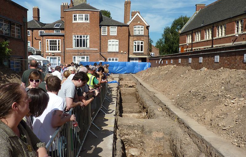
But members of the Richard III Society, the University of Leicester Archeology Department and the Leicester City Council undertook to find Richard’s remains. On August 25, 2012 it was announced that three trenches had been dug under a municipal parking lot, uncovering not only the original walls from the cloister and church, but a single set of human remains buried near the altar. But are these Richard’s remains?

At this point, forensic anthropologists have collected strong circumstantial evidence. The remains belong to a battle-scarred adult man, seemingly buried without clothes, with an arrowhead lodged between two vertebrae and a horrible gash in the skull. But most telling is the curvature of the spine from scoliosis which would have resulted in the living man standing with one shoulder higher than the other. Richard III was rumoured to be a hunchback (as portrayed by Shakespeare) but it is unlikely such an accomplished soldier would have such a severe disability. But a mild curvature of the spine might account for the seed of what later became legend.

Forensic anthropologists hope to definitively identify the remains using mitrochondrial DNA, a type of DNA passed down through the generations via the maternal bloodline. Three Canadians from London, Ontario are the only remaining relatives of Richard III through the maternal line and their DNA will be tested against samples taken from the femur and molars of the remains. Scientists are hopeful of a match, but the age of the samples and the resulting quality of the DNA is a concern. They hope to have a definitive answer within the next three months.

Next week, in our next Forensics 101 post, we’re going to examine how mitrochondrial DNA can be used to identify the dead by comparing their sequences to those of their living relatives.

Photo credit: Wikimedia Commons