Forensics 101: Forensic Challenges of Mass Grave Excavations

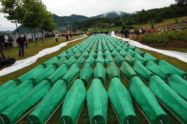
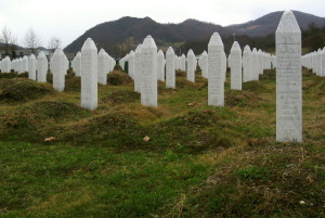
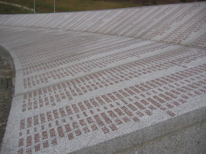
Last week we marked the 18th anniversary of the massacre of 8,100 Bosniak men and boys in Srebrenica by the Bosnian Serbs. The overwhelming majority of these victims were buried in mass graves in the remote countryside. The task for investigators following the massacre was not only finding the gravesites, but successfully excavating and identifying the victims.

The UN defines a mass grave as a location containing three or more victims who have died by extra-judicial or arbitrary executions that are not the result of an armed conflict (an extra-judicial action is one that takes place by a state or other official authority without legal process or the permission of a court).

Investigators need to determine not only time since death, but also discover any evidence of torture, the specific method of death, and the identity of the victim where possible. For many bodies, this may be a near impossible task.

Among the numerous challenges confronting researchers during mass grave excavations in Bosnia were:

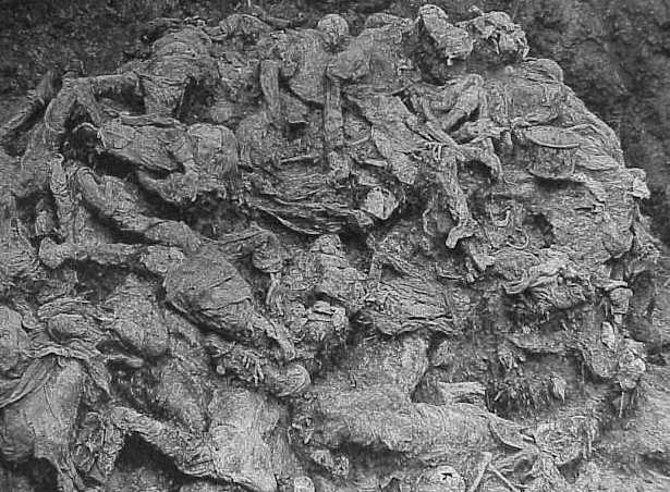
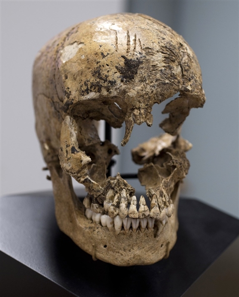
  • State of the remains: Victims were often not buried immediately after death because of the need to bring in heavy equipment to dig the grave. As a result, partially decomposed remains became separated and scattered within a single gravesite. The heavy machinery used to dig mass graves and to transport and bury the dead also caused damage to both the soft tissue and the skeleton, masking original trauma and complicating the investigation.
  • Victim collection and labeling: During any forensic recovery, each separate body part is identified as an individual specimen. Any possible personal effects or related body parts must be labeled with related information for later association, leading to an incredibly complex identification scheme.
  • Secondary and tertiary graves: A large majority of the mass graves in Bosnia were reopened, and disinterred victims moved to secondary or even tertiary graves. Since this occurred anywhere from one and four months post-mortem, soft tissue degradation was well advanced, leading to significant scattering of victims’ remains across large swathes of countryside.
  • Lack of associated physical objects: Bodies were carelessly dumped into mass graves and often tightly packed to keep the site as small as possible. When personal effects were recovered, it was often impossible to determine to whom they belonged.
  • Clandestine sites: Mass graves, by design, were purposely situated in difficult-to-identify locations, usually in remote areas. In addition, the killers deliberately tried to make victim ID difficult by having their victims remove all personal effects, such as wallets and jewelry, before execution.
  • Sheer number of victims: Some mass graves in Bosnia contained up to 700 victims. This made victim recovery and identification a substantial task simply from a procedural and practical standpoint.
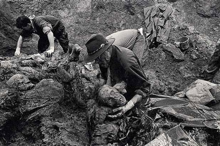
  • Need for large international teams: Human rights horrors such as mass graves are very difficult tasks for investigators, frequently leading to depression and fatigue. Regular replacements are required, and the specialized nature of the work involved requires an international effort to staff a large team. It will normally take 1 or 2 investigators approximately 4 days to excavate a single victim. If a grave has hundreds of victims, it can take a team of several dozen investigators months to complete.
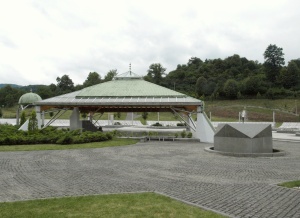
  • Need for on-site facilities: Due to the remote nature of most mass graves, investigators must build or acquire forensic facilities for their investigation—including refrigerated storage areas, running water, decontamination areas, and sorting areas for both remains and personal effects. Provision must also be made for site security during the excavation, and accommodations for the technical staff.
  • Victim identification: The majority of mass grave victims frequently lacked sufficient dental records to allow for dental identification. As a result, pathologists and forensic anthropologists had to rely on physical features and antemortem fractures to establish victim identification.

Next week we’re going to look at the practical side of mass grave excavations—how to find the graves—and then, once they are located, how to recover the victims.

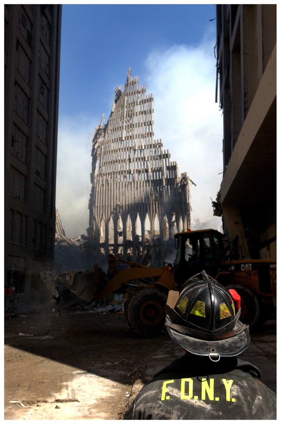
Photo credit: Wikimedia Commons and Gilles Peress.

Forensic Case Files: The Srebrenica Massacre

July 11th this past week marked the 18th anniversary of the beginning of the Srebrenica massacre—the day the Bosnian Serb army, under the command of General Ratko Mladić, took control of the UN protected enclave of Srebrenica in Bosnia.  Two days later the genocide began.  Between July 13th and 22nd, 1995, over 8,100 Bosnian Muslim men and boys were massacred and buried in mass graves by the Serb Army. Between August and November of 1995, many of those bodies were moved to secondary and tertiary mass graves, scattering remains across 300+ grave sites. The locations of these graves were largely unknown to outside investigators, and while a large number of them have been discovered, many are still unidentified.  Teams of pathologists and forensic anthropologists are sponsored by the International Commission on Missing Persons to excavate each newly discovered grave. Attempts are made to identify remains by PCR, physical characteristics and personal belongings found within the grave.  It is truly horrifying work for the ICMP team members, but it is also rewarding as missing loved ones are finally identified and put to rest.

DNA analysis comparing family member samples to the unidentified remains has resulted in the identification of 6,838 individuals from the more than 8,100 reported missing following those 10 days in July. But there remains no trace of over 1,200 men and boys to this day.

On July 11th of each year, all of the newly identified dead are brought to the Srebrenica Genocide Memorial in Potočari for burial.  Last Thursday, 409 additional sets of remains—often no more than a handful of bones—were laid to rest at the memorial. Included in the dead were 43 boys between the ages of 14 and 18, and a newborn infant who was born during the massacre.  This brings the total number of remains interred here to 6,066.

Next week, as we explore this difficult topic further, we’ll look at the forensic anthropology challenges of mass graves.

Photo credit: green-draped coffins—Almir Dzanovic, mass grave exhumation Photograph provided courtesy of the ICTY, Potocari gravestones— Michael Büker, Potocari Memorial—Mazbln and Potocari Memorial names— Michael Büker; all Wikimedia Commons

Forensic Case Files: Guatemalan Genocide

Guatemala was once home to an advanced Mayan civilization that flourished from about 250 C.E. to nearly 1000 C.E. Their declining civilization was overrun by the invading Spanish in the 16th century. Conquered by weapons of modern warfare and devastated by European diseases, the Mayans became the Guatemalan peasant and labouring class to the Spanish upper class. But not even invading Europeans would wreak as much death and destruction as Guatemala’s own government in eighteen months between March 1982 and August 1983.

Coffee production is one of the Guatemala’s main industries and was heavily invested in by Americans during its infancy in the early- to mid-20th century. Large coffee plantations were run by the white upper class, while the indigenous Mayan population worked the fields. As a result, a large gap formed between the police-protected white populace and impoverished natives.

During the 1940s and into the 1950s, successive governments made great strides in improving conditions for the native populations, but a C.I.A.-facilitated coup in 1954 overthrew the existing government due to the rumoured threat of Communism. A military dictator was installed to lead the country and this became the style of government for the next several decades. During that time, several guerilla factions rose up to threaten the government, leading to the Guatemalan Civil War (1960 – 1996). The government’s response was to deal quickly and violently to any guerilla threat.

In March of 1982, General Efrain Ríos Montt overthrew the government in power and installed himself as President. His views regarding the guerilla resistance were very clear: “If you are with us, we’ll feed you; if not, we’ll kill you.” Officially, he ordered paramilitary ‘death squads’ out into the mountains with the intent of discovering and killing guerilla soldiers.

But something much more tragic took place. Over the course of the next year and a half, 669 massacres occurred at Mayan villages. Death squad soldiers would wait until the village gathered together for a celebration or market day, and then the entire community was targeted under the guise of harboring guerilla rebels. Peasants were shot, stabbed or bludgeoned to death. Many had their limbs amputated. Some were impaled and left to die slowly, or doused with gasoline and set afire. Women and girls were raped, the elderly were slaughtered, babies’ heads were smashed against poles, and children were thrown into mass grave pits of the dead and buried alive. Afterwards, soldiers took or killed the livestock, destroyed crops, fouled the local water supply, and desecrated any sacred places. Then they burnt what was left of the village to the ground. It was true ‘scorched earth’ warfare. Those fortunate enough to flee to the mountains were hunted by soldiers with the goal of exterminating the entire village. Hundreds of thousands of displaced peasants became refugees.

More than 200,000 native Mayans were murdered and another 50,000 ‘disappeared’ during that eighteen month period. The Mayan population refers to this time as the ‘Silent Holocaust’—when villagers were killed simply due to their ethnicity, not because they supported any rebel faction. The government supported their actions with the claim that the Mayan communities had organized, allied with the guerillas, and were working towards a Communist coup.

In 1994, FAFG, the Guatemalan Forensic Anthropology Foundation started out as a small group of forensic anthropologists and scientists dedicated to the goal of uncovering the dead from this atrocity. In 1995, those five scientists began their first exhumation aided by massacre survivors. Currently the group numbers more than ninety and FAFG scientists are considered to be the world’s experts on mass graves. They have assisted in exposing other massacres, such as Srebrenica following the Bosnian War. They work to discover and exhume mass graves, recover human remains, determine the traumatic cause of death, and attempt to ID the victims based on skeletal structure and associated grave goods.

Shortly after the exhumations began, the U.N. investigated the Guatemalan genocide. In 1999, they finally released a report detailing horrific human rights violations by the military as ordered from the highest levels of the Guatemalan government.

In 2009, the National Security Archive presented a report citing President Montt and his military of carrying out genocidal assault against the indigenous Mayan population. Part of their supporting evidence was a ‘death squad diary’, outlining the disappearances, tortures, and executions starting in the summer of 1982 and continuing into 1983.

For the very first time, a previous head of state is on trial by the justice system of his own country for crimes committed within that state. Currently 86 years of age, and decades after his time in office, Efrain Ríos Montt now stands trial. So far, more than 70 witnesses for the prosecution have testified to the atrocities.

Unfortunately, the trial has been plagued by procedural and technical errors. On May 10, 2013, Montt was convicted of ordering the deaths of 1,771 Mayan peasants and sentenced to 80 years in prison.  But on May 20, that ruling was overturned based on ‘illegal proceedings’—Montt had fired his attorneys on April 19th and was left without a lawyer for a short period of time while the trial proceeded. Guatemala’s constitutional court ruled this past week that the trial should have been halted until Montt had lawyers in place and that all court proceedings must roll back to April 19th and start again. All witness testimony up to that time will stand, but the final weeks of the trial now must be repeated.

Guatemala stands as the only modern genocide in the Western Hemisphere during the post-World War II era. One can only hope that justice will be done, but it will be a small consolation to the Guatemalan people, many of whom still mourn those lost during the massacres.

Photo credit: Wikimedia Commons

Forensic Case Files: Cannibalism in Jamestown in the Early 17th Century

Jamestown, Virginia was settled on a swampy peninsula in 1607, making it America’s first permanent English colony. The soggy ground was considered un-farmable by the local Powhatan native tribes, and was rife with malaria-carrying mosquitos. But the lack of local inhabitants and a defensible position—the peninsula is surrounded by two rivers and Chesapeake Bay—made it ideal from an English perspective for the planned location of Fort James.

Initially, interactions between the local Powhatan tribes and the English were good—the natives provided food and hoped to continue to do so in trade for European metal tools. But the English, finding that the land truly wasn’t suitable to farm, especially after 1608’s poor harvest, couldn't produce enough food on their own. They attempted to force the natives to provide more food than they had even for themselves. The resulting conflict led to native raids on the fort and, eventually, to the Anglo-Powhatan War of 1610-1614.

The winter of 1609-1610 was especially brutal, and is referred to as ‘the starving time’ in historical records. The lack of food, ravaging disease, and attacks by the Powhatans led to dire conditions. By the time help finally arrived in May of 1610, only 60 of the original 500 colonists were still alive, and the fort was less of a military installation than a charnel house.

Writings of the time tell of cannibalism in the colony—including a husband who murdered his pregnant wife, and then salted and ate her flesh, a crime for which he was later executed. But no recovered remains provided evidence to support these tales. Until last week.

Archeologists were excavating what was essentially a 17th century garbage heap in the a cellar of a dwelling inside the remains of the fort when they unearthed a human cranium, lower jaw and some shattered leg bones scattered among horse and dog bones. Dr. Doug Owsley, a forensic anthropologist with the Smithsonian Museum of Natural History was called in to examine the remains. Long-time readers of this blog may remember Dr. Owsley as the forensic anthropologist who examined the remains of the unknown Union solider discovered in 2008 at Antietam. For the first time, Dr. Owsley was able to substantiate historical records, describing the discovering as ‘very strong evidence’ of cannibalism.

Kerfs from several tools are clearly visible on the bones of the skull. Chops from a hatchet or axe tentatively score the forehead, and then more substantially mar the back of the head as the attacker gained confidence and used more force. Knife marks on the cheeks and jaw show where muscle was sliced from the bone. The left side of skull is missing—tool marks from a pry bar on the remaining bone attest to the fact that the cranium was shattered when it was forced open to extract the brain. Blessedly,the regular nature of the kerf marks reveal that there was no struggle; most likely, the victim was already dead.

Forensic anthropology reveals clues about the victim of this horrific act—she was young, probably about fourteen years of age from the analysis of the skull, teeth, long bones and from epiphyseal fusion at the knee joint. Strontium analysis of the bone has determined that she grew up in England and arrived in America mere months before her death. She likely died of starvation or sickness in the first months of 1610, and, shortly thereafter, met her final fate at the hands of another colonist. Researchers have christened her ‘Jane’.

Most colonies did not last for even a year in the New World, so, in many ways, Jamestown is a story of persistence and survival during the worst of times. Sadly, it was at the cost of too many lives, some of them lived in desperation as the few remaining colonists struggled to hold on at all costs, buoyed by the faint hope of spring and the sight of a supply ship on the horizon.

Photo credit: Carolyn Kaster/AP

Forensic Case Files: 9/11—Part 3: Challenges in Naming the Dead

Over the past two weeks, we’ve discussed the tragedy of the 9/11 terror attacks from a forensic perspective—how recovery teams worked tirelessly to collect the victim remains once all hope of rescue was exhausted, and how victim identification is established. Sadly, a full 40% of victims from the World Trade Center are still unidentified. Hundreds of thousands of man-hours have gone into the effort, so why has the process of naming the dead proved so difficult?

There were many problems associated with victim identification, especially in the years immediately following the tragedy, including:

  • The sheer number of samples needing to be identified and the amount of data they produced: It is the duty of the Medical Examiner and his staff to identify the dead and issue death certificates. That duty doesn’t change simply because of an overwhelming death toll; each individual still deserves to be named. But because of the nature of death, many victims’ bodies were fragmented, leading to multiple samples from the same individual. Fragmented remains found in the same location may or may not originate from a single victim, so each had to be sampled and analyzed separately. Additionally, personal effects found loosely associated with human remains might not belong to that person, so DNA samples had to be taken from all items. While mass-casualty disasters are not uncommon, the data processing requirements for managing such a large database stretched the technology available to individual laboratories of the time.
  • The size and condition of the samples: Due to the harsh conditions of the site, many samples were so badly degraded that DNA typing wasn’t possible. When samples were found five years after the attack on the roof of the Deutsche Bank Building, most of the bone fragments were less than one sixteenth of an inch in size, minimizing the chances of successful DNA extraction.
  • Weathering/scavenging of samples found years after the tragedy: The Deutsche Bank Building fragments, for example, were subject to years of freezing in winter, and heat and direct sun in summer for five years. Remains in the lower levels of the World Trade Center, among the last to be excavated, were subject to water, fire, crushing, and toxic waste. Remains in the pile sent to Fresh Kills were subject to scavenging by carnivores, birds and insects.
  • Location of the remains: The final resting place of the remains could not be used towards a definitive identification. It might, however, suggest a potential localization—bodies from upper floors may be likely to be less damaged due to the lighter load above them, and fire damaged bodies are more likely to originate from floors near the original crash sites and the ensuing fuel-amplified fire. Additionally, co-mingled remains might be thought to originate from similar areas of the building, if not the same area.

In the years since 9/11, a definitive ID for each victim has proven to be impossible, no matter how much effort has was applied to the task. In the end, at the request of families, 1,616 death certificates were issued without confirmatory identification.

The ultimate question in mass casualty disasters is: when is the project finished? When every victim is identified or when every sample of remains is tested? Sadly, with only 1,119 of 2,753 victims identified, the task of identifying the victims of 9/11 may never officially be complete.

In memory of those lost on 9/11. We will never forget…

Photo credit: WikimediaCommons – U.S. Navy, Wikimedia Commons - U.S. Air Force and Morgan.Davis


ARC Giveaway:

A new Goodreads giveaway for a signed ARC of DEAD, WITHOUT A STONE TO TELL IT runs until April 29th! Enter here: http://www.goodreads.com/giveaway/show/51031-dead-without-a-stone-to-tell-it

Forensic Case Files: 9/11—Part 2: Identifying Human Remains

Comparison points of ridge characteristics for fingerprint analysis.

Last week, we talked about the challenges of handing a mass fatality disaster such as 9/11, including the collection of human remains. This week, we’ll cover how those remains can lead to victim identification.

The path toward identification starts with the type of sample recovered. When the body is intact, presumptive identification can be made via visual ID or by directly associated personal effects (i.e. a driver’s license with matching photo found in the pocket of the victim). Confirmatory identification can then be made using one of several methods, including DNA matching, odontology or fingerprinting.

Sadly, considering the nature of the 9/11 attack on the World Trade Center (WTC), the overwhelming majority of remains could not be identified so easily. Officially, the New York Medical Examiner lists all of the deaths at WTC that day as ‘homicide due to blunt force trauma.’ This includes those who died in the collapse of the towers, as well as those that fell or jumped to their deaths after being driven out by flame and smoke (these deaths are not classified as suicides since they were not considered voluntary acts).

Because forensic anthropologists specialize in fragmented, burned, decomposed, and comingled remains, they are at the forefront working on victim identification. Well-known author Dr. Kathy Reichs was one of many forensic anthropologists who took time away from their own professional careers to help identify remains found at Ground Zero following the attacks.

For most victims, since only fragments of their bodies were recovered, identification had to be inferred from one or more of the following attributes:

  • Personal surface markers like scars or tattoos.
  • Forensic anthropologists’ estimate of age at time of death, race, sex, and stature.
  • Description of antemortem (before death) characteristics, including evidence of disease or healed fractures.
  • Discovery of prosthetics or surgical hardware (including serial numbers).
  • Documentation of perimortem (at the time of death) trauma supporting cause of death.
  • Fingerprint examination: Qualified personnel can collect antemortem latent prints from the homes or personal effects of suspected victims for comparison to recovered remains. Once identification is made, a second qualified examiner must confirm the match.
  • Odontology: Comparison of recovered dental fragments to antemortem dental x-rays and charts. These matches can be difficult because dental remains may be fragmented; extremely fragile dental remains may require onsite radiography before transportation to morgue.
  • Radiology: Comparison of antemortem x-rays to post-mortem (after death) x-rays and skeletal fragments in order to match healed fractures.
  • DNA comparisons: DNA remains the best method of identification, especially when other physical traits such as fingerprints, physical stature, distinctive characteristics and dental features have been destroyed. The challenge in DNA matching can lie in finding a reference sample for comparison. More detailed information on the subject can be found in one of our earlier posts: Forensics 101: DNA Profiling for Identification.

In a perfect world, every victim would be identified, finally bringing closure to the families. But the task of identifying the victims at the WTC has proven to be extremely difficult in many cases. Join us next week as we close our series on 9/11 as we explore the challenges investigators have faced in trying to put names to the dead.

Photo credit: Vince Alongi


ARC Giveaway!

A new Goodreads giveaway for a signed ARC of DEAD, WITHOUT A STONE TO TELL IT runs until April 19th! Enter here: http://www.goodreads.com/giveaway/show/49884-dead-without-a-stone-to-tell-it

Forensic Case Files: 9/11—Part 1: Mass Fatality Incidents

The events of September 11, 2001 will forever remain a watershed moment in history—life before vesus life after that day. For North Americans, it marked the end of a more relaxed way of life and the beginning of heightened security and wariness of the world around us.  For most of us, it’s an event, like Kennedy’s assassination, that will forever be linked to what we were doing at the moment we heard the news.

In the past, Ann and I have considered looking at the recovery efforts assocated with the disaster because forensic anthropology remains a crucial part of victim identification to this day. But, at the same time, we’re very sensitive to the fact that this incident remains a very painful moment in time not just for Americans, but for the world as a whole since sixty other countries also lost citizens in the attack that day. Over the next few weeks, we’re going to be looking at the incident from the perspective of managing a mass casualty and fatality incident of this magnitude, and continuing efforts at individual victim identification.

When the planes struck the two towers, significant damage was initially localized to seven or eight stories adjacent to the point of impact, caused by explosion, fire from the heavy load of airplane fuel, and the large size of the modern Boeing 767. The buildings’ collapse was initiated by the weakening and finally buckling structural systems due to the heat of the fire and the crushing static weight of the floors above. The South tower, the second hit, was actually the first to collapse because the plane struck a lower floor, resulting in greater weight above the site of impact.

The sheer volume of calls overloaded communications systems, making it difficult to contact those inside the buildings, including first responders. As a result, many in the North Tower were never aware that the South Tower had fallen, even though nearly thirty minutes passed before the North Tower itself collapsed. 2,753 people, including the passengers and crew of American Airlines Flight 11 and United Airlines Flight 175, perished in the tragedy.

The initial response was search and rescue in an attempt to recover anyone who might have survived the crushing collapse of either building. The instability of both the immediate scene and the surrounding buildings hampered rescue attempts and teams were called off repeatedly as concerns about the collapse of nearby buildings heightened; 7 World Trade Center collapsed later that afternoon as a result of the fires that started after the building was hit by debris from the North Tower. Only when the scene was stabilized were rescue workers allowed to return. Multiple hazards were also a concern throughout this phase, including an underground tank of diesel fuel, gasoline from several thousand cars buried in the underground parking lots, and 1.2 million rounds of ammunition in the U.S. Customs Service firing range on site. Sadly, in the days following the attack, only 11 survivors were pulled from the rubble. Some victims survived the collapse of the towers but rescue teams were unable to reach them in time.

Recovery teams formed bucket brigades, passing five-gallon buckets down lines to investigators who sifted through each to remove any evidence of human remains. ‘The Pile’ was then transferred to one of several landfill sites, including Fresh Kills on Staten Island. There, the debris was sorted once again, and any additional human remains and personal effects were collected. The majority of remains collected were recovered during the ten months following September 2001.

Salem Fire Department’s 9/11 memorial, including a steel girder from one of the towers.The New York City Office of Emergency Management was in charge of the recovery and cleanup. Keenly aware of the effect on the city of the specter of the wreck of the World Trade Center, they attempted to clean up the 130,000 tons of debris as quickly as possible. Inadvertently, this rapid cleanup caused some remains to be separated from personal effects which could be used to aid in victim identification, and further scattered the remains of dismembered bodies. Inadvertently, human remains may have been disturbed as remains and comingled effects became separated, or as associated remains became scattered. In 2005, the search was declared complete despite concerns raised by families of those still missing that the initial efforts had been too rushed or carelessly handled. But after the discovery of bone fragments on the roof of the nearby Deutsche Bank Building and in two manholes in 2006, a new investigation was launched and 1,500 additional remains were recovered.

Twelve years after the attack, the cleanup process continues. In just the last few years, over sixty truckloads of debris have been removed from the site. On April 1, 2013 two more skeletal fragments were discovered. Currently, 40% of the victims are still unidentified, so efforts to identify the missing and the dead will continue.

Next week, we’ll look at identification methods used following the attacks to identify the dead.


ARC Giveaway!

A new Goodreads giveaway for a signed ARC of DEAD, WITHOUT A STONE TO TELL IT runs until April 19th! Enter here: http://www.goodreads.com/giveaway/show/49884-dead-without-a-stone-to-tell-it


Forensics 101: Bullet Wounds in Bone—The Skull

In a previous Forensics 101 post, we looked at how kerfs—the grooves and notches made by tools on bone—can help scientists identify the method of death in a murder investigation. But the rise of gun crime in North America has made the forensics of wound ballistics increasingly important. There are two different types of damage in this kind of wound—soft tissue and bone. In this post we’re going to strictly look at bone damage, concentrating on the skull and its very characteristic fracture patterns.

Unlike blunt force trauma, gunshot wounds often cause both an entrance and an exit wound. Investigators need to be familiar with how bone behaves in both circumstances to reconstruct the order of events and be able to piece together the details of the fatal shot. Different variables that affect the type of damage done to the bone include the velocity of the bullet (which depends on the type of gun used and the distance between the shooter and the victim), the size/caliber of the bullet, and the angle of impact.

If a bullet penetrates the skull perpendicular to the surface, a round defect is formed, often with outward radiating fractures extending from the bullet hole. The force of the bullet’s entrance increases the intracranial pressure inside the skull, causing the pieces of bone between the radiating fractures to push outwards. These ‘heaving fractures’ can be differentiated from blunt force trauma fractures because the bone sits above the plane of the skull instead of below it. The energy transfer from the bullet to the bone can be so efficient that the radiating fractures can travel through the bone to the far side of the skull faster than the bullet can traverse the brain and exit. This fact can be crucial in determining the order of fractures since a new exit fracture cannot cross an existing entrance fracture.

When a bullet strikes the skull tangentially, a characteristic ‘keyhole’ is formed—a defect that is circular at one end with tangential fractures radiating outwards in parallel, allowing the bone between them to lever out.

Exit wounds often tend to be much larger than entrance wounds for a number of reasons: the bullet is misshapen or ‘mushroomed’ from the initial bone strike, the bullet may no longer be moving along a straight trajectory, or the projectile may be tumbling end-over-end. Often large chunks of bone may be completely detached from the skull following the bullet’s exit. Sometimes, however, the bullet’s energy is spent following the initial strike; when this occurs, the bullet does not exit the skull and can be recovered later during autopsy.

Contrary to popular belief, the size of the bullet wound does not directly correlate to an exact bullet caliber because factors such as bullet shape, jacket material, stability of the bullet’s flight path and whether any other targets have been hit tangentially can affect the force with which the bullet strikes the bone.

Photo credit: Ann H. Ross, The University of Tennessee and Gérald Quatrehomme et al, Florida Atlantic University

ARC Giveaway!

A new Goodreads giveaway for a signed ARC of DEAD, WITHOUT A STONE TO TELL IT runs until April 8th! Enter here: http://bit.ly/10ghlSr

Forensic Case Files: Black Death Victims Uncovered in London

68216_farringon_charterhouse_square_archaeology_march_2013.jpg

A burial ground containing thirteen sets of remains was recently discovered in London during excavations for the Crossrail project, the expansion of London’s existing public transit rail lines. Located in Farringdon—an area of downtown London—in one of the rare undeveloped sections of the city, the remains were uncovered in a location suspected to be one of the city’s emergency burial grounds used during the 14th century plague known as the ‘Black Death’. Open in 1349 and possibly receiving up to 50,000 dead during the next three years, the Farringdon burial ground was referred to in historical texts as ‘no man’s land’. Closed in the 1500’s, its exact location was lost to time until now.

Laying a mere eight feet underground, the skeletons were discovered in two neat rows. The burial pattern indicates that this area of the cemetery was used at the beginning of the plague, when death rates were low and individual burials were common. In later years, the overburdened and fearful population simply discarded the constant stream of bodies into mass graves. An indication of the mindset of the population at the time—in 1347, the average number of wills registered in London was only 20; but by 1349, that number ballooned to 370. Simply put, the English expected to die, and wanted no contact with the sick or the dying that lessened their chances of survival.

Osteologists and archeologists from the Museum of London are already excavating and removing the remains for study. They plan to extract plague DNA from the tooth pulp of the victims and hope to be able to sequence the bacteria’s genomic DNA, possibly mapping it as the ‘mother of all modern plague species’. The remains will also give scientists a glimpse into life in the 14th century—the wear patterns on the bones reveal it was a life of heavy labour, but other indicators will give information about their general health and stature.

Black Plaque woodcut.png

Yersinia pestis, the bacteria responsible for the Black Plague, originated in China and was brought to Europe in the mid-14th century. It started in Persia in 1346 and then spread through southern Europe in 1347 before moving north into the rest of Europe and Russia over the next five years. It arrived in London and the south of Britain in 1348 before spreading to the rest of the country and beyond. By the time the epidemic played out, an estimated 75 – 200 million people, roughly 30 – 60% of Europe’s population were dead. Carried by rats, and transmitted by the rodents’ fleas, Y. pestis sickens it’s victims by suppressing the body’s normal immune response. It directly affects immune cells in the blood and evades the body’s response by hiding and replicating in the lymph nodes, creating the characteristic black buboes or swollen lymph nodes that often oozed pus and blood. Medieval physicians often lanced the buboes, exposing the unsuspecting practitioner to the infectious contents and spreading the plague further. The number of infections dramatically decreased in Britain after 1350, but smaller outbreaks continued for the rest of the 14th century. A second major plague took place between 1665 and 1666 in London, but by the late 18th century, it had mostly disappeared from Europe.

An electron micrograph of a cluster of Yersinia pestis bacteria.

An electron micrograph of a cluster of Yersinia pestis bacteria.

Why the plague died out has never been definitively answered, but there are several possibilities. Those that survived the first wave of the plague in 1348 became immune to further infections, leaving fewer susceptible to future infection. The 1348 wave of the plague mostly infected adults, but later waves—1361 for instance—primarily infected children who were not alive in 1348 and had no resistance to the bacteria. Later on, herd immunity (the greater immune population protecting the lesser susceptible population simply by their inability to become infected) would have protected younger members of the population. There is also a theory that the fleas that carried Yersinia pestis only lived on black rats. When larger brown rats out-competed the black rats in Europe, there were fewer carriers for the plague.

Photo credit: Crossrail, Rocky Mountain Laboratories/Wikimedia Commons, and the Toggenburg Bible.

Giveaways!

It’s the final days for Five Star’s Goodreads giveaway! 10 ARCs of DEAD, WITHOUT A STONE TO TELL IT are up for grabs to American entrants, ending on Sunday night: http://www.goodreads.com/giveaway/show/41803-dead-without-a-stone-to-tell-it

And stay tuned for more giveaways coming soon!

Forensics 101: Using the Bomb Curve to Date Human Remains

Ivy_Mike_-_mushroom_cloud by FastFission.jpg

Over the past month, we’ve discussed human remains that were centuries—King Richard III—if not a millennium old—King Alfred the Great. For remains of this age, classic carbon dating is the most reliable way of determining time since death. But is there a more precise way to date more recent remains, remains that might only be thirty to fifty years old, instead of six hundred? There is, and that method uses the fallout from nuclear testing following the Second World War to determine time since death.

Following the end of the Second World War, nuclear weapons were tested by the United States, the United Kingdom and Russia. The fallout from this testing radically changed the percentage of radioactive carbon—14C—in the atmosphere, spiking significantly in the early 1960’s before peaking in 1963 at a level nearly twice that of 1950. Atmospheric 14C levels fell slowly in the decades following, but still remain 15% higher than in 1950.

Average atmospheric 14CO2 for the northern hemisphere

Average atmospheric 14CO2 for the northern hemisphere

Just as strontium is incorporated into living organisms, 14C in atmospheric CO2 enters the food chain when plants use it to manufacture carbohydrates and proteins during photosynthesis. Those plants are then eaten by herbivores and become a permanent part of that animal’s bone structure. As a result, 14C from samples taken from skeletal remains after the 1950’s can be compared to the bomb curve to determine relevant dates. Samples taken from the mid-shaft of long bones represent childhood 14C levels. Spongy cancellous bone sampled from the ends of long bones will show a greater amount of turnover and remodeling that correlates closely to the date of death. Enamel from teeth captures a snapshot of the time when the tooth developed and erupted. If all the values fall in the pre-1950’s range, a different manner of aging the remains must to be used. But for those values that fall post 1950, a window of only a few years can be determined for the date of death.

The slow drop in atmospheric 14CO2 following the early 1960s is due to the signing of the Limited Test Ban Treaty. In August of 1963, representatives from the United States, Russia and the United Kingdom signed a treaty banning all nuclear testing in the atmosphere, in space or under water. In the decades that followed, 123 additional countries signed the ban (the most recent was Montenegro in 2006), leaving 58 states as non-signatory.

Photo credit: Fastfission via Wikimedia Commons and Ubelaker, DH et al. Analysis of Artificial Radiocarbon in Different Skeletal and Dental Tissue Types to Evaluate Date of Death. Journal of Forensic Sciences; May, 2006

Giveaways!

A new Goodreads giveaway starts today! I’m giving away an autographed ARC to Canadian entrants. The contest closes on March 15, 2013: http://www.goodreads.com/giveaway/show/46552-dead-without-a-stone-to-tell-it

Five Star is giving away 10 copies to American entrants, ending on March 25, 2013: http://www.goodreads.com/giveaway/show/41803-dead-without-a-stone-to-tell-it

A Facebook giveaway! Stop by Facebook for your chance to enter a Rafflecopter giveaway for a chance to win an autographed ARC: http://is.gd/iDxH1z

Forensic Case Files: 9th Century Remains of King Alfred Discovered?

It seems like English kings are popping up all over the place recently. A few weeks ago I shared the story of King Richard III’s positively identified remains in Leicester. Shortly after that story broke, Ann (who is always my best resource) found a story about archeologists in Winchester who believed remains found in an unmarked grave are those of King Alfred the Great (849 – 899 A.D.).

Following the death of his three older brothers, Alfred took the throne at twenty-two. Over the course of his reign, he became best known for keeping the Viking invasion at bay, thus ensuring English as the country’s spoken language and Christianity as its religion. First called the King of Wessex (an area of southwestern England), he was successful at repelling the Danish invaders in his own kingdom before recapturing London from the Danes and making peace with their leader, Guthrum. Known afterwards as the first King of the English, Alfred was also responsible for introducing new military responses to specifically counter new Danish strategies, significantly improving England’s naval power, introducing the country’s first written code of law, and introducing a standard system of coins.

Following his death in 899 A.D. from natural causes (possibly Crohn’s Disease), Alfred was buried in first in Winchester, a city in the south of England. Later, his remains were transferred to the newly opened Hyde Abbey in 1110, along with the remains of his wife and children. In 1539, during Henry VIII's dissolution of all Catholic abbeys and monasteries, Hyde Abbey was demolished, but the graves were left intact. In the eighteenth century, the graves were robbed and stripped of all contents, and later excavations showed Alfred’s grave to be empty.

In the late 1800’s, a vicar at St. Bartholomew’s Church in Winchester paid ten shillings for a collection of bones rumoured to have originated at Hyde Abbey. These bones, including five skulls, where interred at St. Bartholomew’s Church until archeologists recently identified the remains as possibly belonging to King Alfred the Great.

This archaeological team will face challenges that Richard III’s team did not, including:

  • Multiple individuals interred in the same grave—a forensic anthropologist will be able to determine sex from some bones, relative height from others, and will try to separate each individual’s remains.
  • Age of the remains—King Alfred’s remains are six centuries older than those of Richard III, and the chances of extracting viable DNA are much smaller. Scientists have been trying to recover DNA from Alfred’s granddaughter's remains, but have not yet been successful.
  • No known living relative—Genealogy before the turn of the first millennium was not documented as it was after Richard III. If Alfred’s family line continues to this day, his relatives are most likely unaware of the connection.
  • Radiocarbon dating is probably the best chance to age match the remains.

The team will begin excavating the remains during spring 2013 and hopes to have results by summer.

It's Goodreads giveaway time! Five Star is giving away 10 copies to American entrants, ending on March 25, 2013: http://www.goodreads.com/giveaway/show/41803-dead-without-a-stone-to-tell-it

Photo credit: Odejea via Wikimedia Commons

Forensic Case Files: Richard III’s Remains Confirmed

On August 25, 2012, the Richard III Society, the University of Leicester Archeology Department and the Leicester City Council made an astounding announcement. Their joint efforts to find the remains of King Richard III had led them to a local municipal parking lot. The three trenches dug there not only revealed the walls from the Greyfriars Church in which Richard was said to be buried, but also a set of human remains—remains with a curious curvature to the spine and signs of violent, battle-related death. They proposed that this skeleton was the remains of Richard III, the last English monarch to die in battle, but they needed time to definitively prove his identification.

We covered this story last fall when the remains were finally excavated, and in the following posts concerning Richard’s supposed (and unproven) treachery in killing his nephews, the Princes in the Tower, and outlining the science that could ultimately prove Richard’s identity.

Yesterday, it was announced that ‘beyond a reasonable doubt’, the remains recovered were indeed those of King Richard III. It took the team over four months of intense testing using the tools of DNA analysis, forensic anthropological examination, carbon dating and environmental analysis to make this determination.

This is the evidence to support Richard’s identification:

  • Despite fears that nucleic acids within the bones might be too degraded after more than 500 years in the ground, DNA was successfully recovered from the teeth. Independent testing in Leicester and York confirmed the mitochondrial DNA match between the remains and Michael Ibsen of London, Ontario, a direct descendant of Anne of York, sister of the king. A second descendant was also found; this too was a match.
  • Carbon dating placed the remains between 1450 and 1540. Richard died at the Battle of Bosworth Field in 1485.
  • Examination of the remains by a forensic anthropologist determined them to be from an individual in his late twenties or early thirties. At the time of his death, Richard was 32.
  • Radiocarbon dating revealed that the individual had consumed a high protein diet. Meat consumption in the fifteenth century was rare, except by those of high social status. 
  • There were ten injuries to the skull, including two fatal wounds by a sword and a halberd (a pole topped by a spiked axe). The latter removed a large chunk of the back of the skull.
  • The remains showed signs of severe scoliosis, which would present as the right shoulder sitting higher than the left. However, both arms appeared normal, in opposition to the Tudor portrayal of Richard as a ‘hunchback with a withered arm’.
  • Body positioning of the remains suggests that the hands were tied, as a prisoners’ might be.
  • The body was interred in a hastily dug grave. It was not long enough, causing the head to tip up so the body could fit. There was also no shroud or any grave artifacts. This careless treatment suggests burial at the hands of the victors of the battle and not those loyal to the Plantagenet cause or its king.

Richard’s remains will be re-interred at Leicester Cathedral. Next up for the Richard III society is to restore Richard’s tarnished reputation. Is it all a result of the victorious Tudor’s and their re-telling of history, or was any of it truth? This will be a challenge as they have centuries of belief to overcome.

For anyone wanting more information about Richard, the search for his remains and how they confirmed his identity, the University of Leicester has set up an excellent website containing all the information they now know: The Search for Richard III – Completed

Photo credit: The University of Leicester

It's Goodreads giveaway time! Win an ARC of DEAD, WITHOUT A STONE TO TELL IT in one of two ways:

1) Five Star is giving away 10 copies to American entrants, ending on March 25, 2013: http://www.goodreads.com/giveaway/show/41803-dead-without-a-stone-to-tell-it

2) I'm giving away an authorgraphed ARC to Canadian entrants starting today and going until February 18th (with more North American giveaways coming soon): http://www.goodreads.com/giveaway/show/44027-dead-without-a-stone-to-tell-it

Forensics 101: Forensic Entomology

Determining time since death of a body more than a few days old can be problematic for investigators. They have a number of tools at their disposal—for example, the stage of decomposition/advance decay, or soil analysis to determine the extent of the body’s chemical breakdown—but the science of entomology can be a more precise way to determine when death occurred. Using knowledge of the life cycle of local carrion insects, scientists can accurately estimate a minimum time since death from one day to more than one month.

Flies:

  • Flies are the first responders of the forensic entomological world, including species such as blow flies (Calliphoridae), muscid flies (Muscidae), and flesh flies (Sarcophagidae).
  • Flies are capable of burrowing and colonizing bodies buried up to 30 – 50 cm deep.
  • Blow flies are usually the first to arrive, being able to detect a body at distances up to one mile. Adult flies can arrive to colonize a corpse within minutes.
  • Most adult flies oviposit (lay eggs) on the body, preferring the moist mucous membrane openings of the face. They are also attracted to any areas of trauma; in fact, this specific colonization indicates areas of damage to the medical examiner before an autopsy is started.
  • Sarcophagidae is the exception to egg laying. It deposits larvae directly onto the corpse.
  • Fly eggs hatch within 8 – 24 hours, and the resulting larvae or maggots will feed off the corpse. A heavily colonized corpse can be completely reduced to skeleton in only a matter of weeks by nothing more than extremely active maggots during warm weather.
  • When a victim is discovered colonized by maggots, investigators will collect samples. Some larvae are sacrificed for DNA which will be analyzed to determine their species. Others will be raised in the laboratory, and scientists can observe them to determine when they pupate and develop into the adult form (this will also confirm or determine species identification). Using knowledge of the species’ life cycle, scientists will then be able to work backward to determine when eggs were laid on the corpse. This is the minimum possible time since death. The maximum time since death estimate is often dependent on the condition of the victim ie. body location, or a time of year that might slow near-instantaneous insect colonization.
  • A body undergoing decomposition moves through the stages of fresh, bloated, decay and dry. Since ovipositing flies require a moist environment for egg development, most flies are no longer attracted to the corpse by mid-way through the decay stage

Beetles:

  • Beetles tend to arrive following the fresh body stage. They will feed on any young fly larvae present as well as the flesh of the corpse itself.
  • Adult beetles will lay their eggs on the corpse; when the larvae hatch, they will consume the corpse as their sole food source.
  • As the body decomposes and dehydrates, beetles remain, feeding on the dry tissue, hair, and any fungus growing on the flesh.
  • As with flies, investigators can collect beetle larvae and hand-raise them in the lab to determine both species and a minimum time since death based on knowledge of the species’ life cycle.

The absence of carrion insects is also important information for investigators. For instance, if a decomposing body lacking in larvae was found in a wooded area in a temperate climate, investigators would likely suspect that the body had only recently been placed there after being stored indoors and out of the contact with insects normally plentiful in that environment. Conversely, if a body was placed outdoors during the winter and mummified due to cold temperatures and low humidity, once insect activity resumed with warmer weather in the spring, flies would never colonize and only beetles would be attracted to the remains.

Using knowledge of the life cycle of key insect species, scientists can determine time since death on older remains. With this information, criminal investigators can begin their investigation to determine the suspect responsible for the death in question.

Photo credit: Wikimedia Commons – Cynomya mortuorum and Philonthus

Forensics 101: Tool Marks in Bone

When skeletonized human remains are recovered, sometimes the only evidence police and scientists have to determine cause of death is the bones themselves. The bones are examined to identify any remnants of tool marks—also called kerf marks—that might indicate a traumatic injury. If the body has been dismembered, those kerf marks can lead directly to the tool(s) used postmortem, even if they might not directly suggest a cause of death.

Any sharp implement applied to bone with sufficient force will leave a distinctive imprint, be it scavenger teeth or a cutting tool. Examination of the bone both macroscopically (using the naked eye) and microscopically (using a light microscope/scanning electron microscope for magnification) can provide crucial information, since each tool leaves a characteristic mark that can assist in its identification.

Types of cutting tools:

Knives: Knives are narrow bladed and leave a corresponding narrow ‘V’-shaped trough in the bone. They are single or double-bladed (double-bladed knives can leave an opposite ‘V’-shaped trough in surrounding bone) and tend to leave behind only microscopic striations.


Saw: Saw blades come in many different form factors, but uniformly leave a wider, square-bottomed trough in bone. They tend to leave distinguishable striations that are easily seen by the naked eye. Individual blade and tooth size can be identified based on the striations, as can the blade type—ie. straight or rotary. Saw kerfs often have characteristic accessory marks, such as false start notches, especially when manual saws are used.


Axe: Like a knife, an axe leaves a smooth ‘V’-shaped trough in the bone, but the defect is very wide and is often significantly deeper due to the lever action of swinging an axe. These kerf marks are often accompanied by microscopic or macroscopic impact fractures and/or flaking surrounding the contact site.


Besides providing information about the type of tool used, kerf marks also provide contextual information, including the handedness of the attacker, the relative positions of attacker and victim, whether the wound was self-inflicted, and the motion of the blade (cut vs. stab). With this information, police can determine not only the most likely weapon used, but how the murder was committed, and these details can often be used to definitely identify the murderer.

Forensic Case Files: The Princes in the Tower

Princes in the tower.jpg

In the last Forensic Case File, I discussed the remains of Richard III, finally discovered in Leicester (pending confirmation). Richard III is infamous for supposedly killing his two nephews, Edward V, 13, and his younger brother Richard, 10, the sons of Edward IV. But because Parliament ruled Edward IV’s marriage invalid and his sons illegitimate, the crown legally went to Richard III in June of 1483. That month, Richard sent his two nephews to the Tower of London. They were seen playing on the grounds over the following months, but were never seen again after the summer of 1483. Surely they had been killed, but the question was by who and when?

The two most famous accounts of the deaths of the young princes were written by Thomas More, Henry VIII’s Lord Chancellor, and William Shakespeare, the famous poet and playwright. Both accounts paint Richard III with a dark brush, portraying him as a villainous monster. But the truth of the matter is that both accounts were written by men who were never witness to the supposed crime—More was eight years old when Richard fell in battle, and Shakespeare was born seventy-nine years later. History tells that More’s account of Richard III came from John Morton, the Archbishop of Canterbury during Henry VII’s reign and a sworn enemy of Richard III. It’s a well-known fact that history is written by the victors; in this case, the House of Tudor was legendary for removing any and all rivals to the throne. It’s not much of a stretch to suppose that the story of Richard III as a menacing hunchback who murdered his nephews was used to blacken the name of a defeated rival family, the House of York.

The truth of the matter is that Richard III had no concrete reason to kill the boys and nothing to gain from their deaths. His claim to the throne came with the approval of both the English public and Parliament. Had Edward IV’s marriage been upheld and his royal line remained intact, there were three other male heirs closer to the throne than Richard, so simply killing his nephews would not have assured him a place as monarch.

So if Richard III wasn’t responsible, who else stood to gain from the princes’ deaths?

In her classic 1951 novel, The Daughter of Time, novelist Josephine Tey makes a convincing argument that Henry Tudor had to most to gain following Richard III’s death at the Battle of Bosworth Field when he was crowned Henry VII.

Henry never missed an opportunity to vilify Richard posthumously, yet when he came to London he never remarked on the fact that the boys were missing from the Tower. It’s been suggested that he would have happily pinned the disappearance and murder of the boys on the fallen king simply to make himself look more favourable by comparison. But perhaps he didn’t want to bring the princes’ disappearance to light because he had ordered their deaths. The boys were also a threat to him—following the death of Richard III, Henry VII repealed the Titulus Regius statute that made Richard III king, nullifying Richard’s reign while simultaneous declaring Edward V King of England and his brother the heir. The boys were a direct threat to his reign as monarch. The boys were never seen after 1483, but it is possible that they were still alive in the Tower as late as 1485 when Henry VII arrived in London. Killing the princes would have ended the last remaining hope that the House of York would be able to reclaim the throne.

Officially, the murder has been laid at the feet of James Tyrrell, an English knight in the service of the House of York and under Richard’s command. Nearly twenty years after the rise of Henry VII, Tyrrell supposedly confessed to smothering the boys and was hanged for his crime. But there was never any transcript of the confession, no formal charges were laid, nor was there ever a trial. In fact, the account was only released after Tyrrell himself was already dead. According to the reported confession, the only witness to the crime conveniently died at the Battle of Bosworth Field along with Richard III.

In 1674, nearly 200 years after the princes’ disappearance, the skeletons of two children were found in a wooden box beneath a stone staircase during renovations in the White Tower. Since it was assumed that these were the remains of the missing princes, the bones were interred with royal honour in an urn in one of the walls of the Henry VII Chapel in Westminster Abbey.

In 1933, Professor William Wright, one of the foremost anatomists of his day, was granted permission to examine the remains. Inside the urn Dr. Wright found the incomplete remains of two children he determined to be between the ages of 7–11 and 11–13 years of age, mixed with several animal bones and a handful of rusty nails. There was no sign of any skeletal trauma on the remains, supporting the theory that the princes had been suffocated. But as the sex could not be determined on the pre-pubescent bones and DNA analysis was decades away, that was the only information learned and the bones were re-interred. Current osteological research now gives scientists methods to determine age of pre-pubescent individuals (although it remains a somewhat inexact science) and DNA analysis could identify the remains if a female family member could be found. Since relatives found to test against Richard III’s remains are not in the same female line as Edward IV’s sons, a new family member in the maternal line would have to be located for confirmation.

So, the mystery surrounding the death and identity of the lost princes remains. If scientists are allowed to examine the remains again, some new answers may come to light. But, unfortunately, it remains a real possibility that the fate of the princes may remain lost to the ages.

Photo credit: Wikimedia Commons

Forensics 101: DNA Profiling for Identification

VNTR gel.gif

Last week, I covered DNA as a tool for identifying remains. This week, I’m going to discuss how scientists test DNA to prove an identifying match.

DNA strands contain different regions, many of which are genes that code for essential protein products. But a very large proportion of sequences don’t code for any currently known genes. These sequences also contain short tandem repeats (STR)—small snippets of DNA that are two to six base pairs long and repeat from three to one hundred times in a row. The locations of these repetitive sections are called variable number tandem repeats (VNTR). Genetically speaking, unrelated individuals will have different numbers of repeated STR segments at known VNTR locations, but related family members will share similar numbers of repeated segments. Since human offspring share a combination of traits from both parents, the power of DNA profiling lies in analyzing numerous segments to definitively prove identification. In North America, it’s standard protocol to analyze thirteen specific locations simultaneously.

Scientists use the polymerase chain reaction (PCR) to examine known VNTR locations. PCR is an assay used to amplify small amounts of specific DNA sequences so they can be visualized later on a gel (we’ll look at PCR in more detail in a future Forensics 101 post). The picture above illustrates typical PCR results. In this case, a gel shows the difference in length of the D1S80 VNTR location of six unrelated individuals, flanked on each side by a marker of known size. As you can see, the pattern for each individual subject is different in each vertical lane.

An example of DNA profiling between a father (1), mother (3) and child (2).

An example of DNA profiling between a father (1), mother (3) and child (2).

There are two types of matches in DNA profiling—identity and inheritance. In identity matching, the unknown sample is tested against a known individual’s DNA. If the two samples match exactly, then the unknown person is identified as the known donor. In inheritance matching, the unknown sample is tested against a sample drawn from a potential family member. If parents are used as donors, then each band in the unknown sample must match with one of the two parents. If a more distant relation is used, then degrees of relatedness are calculated into the expected results.

The bottom line of this testing is that the unknown sample must be tested against a sample (blood, hair, tissue etc.) of known origin. In the case of Richard III’s ancestors, separated by five centuries from the king himself, genomic DNA profiling as described above simply wouldn’t be feasible due to the number of generations separating Richard III and his current ancestors. But when this same method is applied to mitochondrial DNA, because of the consistency of maternal transmission, identical or near identical results are expected between family members, even those separated by multiple generations.

Next week, we’ll look at the historical details surrounding the Princes in the Tower, the young boys Richard III is accused of murdering. Nearly one hundred years after their disappearance, bones were recovered at the Tower of London. But where they of the missing princes? See you next week to find out…

Photo credit: PaleWhaleGale and Magnus Manske

Forensics 101: Tracing Lineage Through Maternal Mitochondrial DNA

DNA-molecule3.jpg

Deoxyribonucleic Acid (DNA)Last week in a post on the potential discovery of Richard III’s remains, I wrote that scientists hope to confirm the identity of the remains based on DNA, specifically mitochondrial DNA. Over the next few weeks, I’m going to explain DNA identification, starting with the basics and then branching out into how it is used to name the dead.

DNA (deoxyribonucleic acid) is the code of life, the genetic information contained in every cell in the form of twenty-three pairs of chromosomes (except for sperm and eggs cells which have twenty-three single chromosomes). The information contained in this genomic DNA specifies everything about us, from how we look to which health problems we’ll have an increased likelihood of developing. But there is also another kind of DNA contained in our cells—mitochondrial DNA (mtDNA).

Mitochondria are the power houses in our cells, the organelles or cellular structures that are responsible for producing the chemical energy required for every cellular activity—from protein production to ion transport to cellular reproduction. But unlike other organelles, mitochondria have their own unique tiny DNA genome—mtDNA. Due to the process of human fertilization, these small bits of DNA are transmitted in family lineages only through the maternal line.

Electron micrograph of two mitochondria

Electron micrograph of two mitochondria

An egg cell is a stripped down version of a typical cell in the body; essentially it only contains a nucleus carrying twenty-three single chromosomes plus several hundred mitochondria in the cytoplasm. A sperm cell is a small sack of DNA attached to a long tail for mobility, the base of which is packed with energy producing mitochondria to fuel the journey. At the moment of fertilization, the tiny sperm head fuses with the egg membrane and injects its DNA while the tail drops off and is lost. As a result, the only DNA inserted into the egg cell is the genomic DNA contained within the sperm head. All mitochondrial DNA that is then replicated as the fertilized zygote splits from one cell into approximately one hundred trillion cells comes solely from the mother. Fortunately, due to the nature of its sequences, mtDNA has a very low mutation rate. In other words, the same mtDNA is passed from grandmother to mother to child through the generations. Genetic testing of specific sequences of those mtDNA samples, even if separated by many generations, can definitively prove a family match.

Due to the number of copies of mitochondrial DNA in each cell (providing approximately five hundred identical copies of each gene versus two copies on the cell’s genomic DNA), forensic anthropologists often are more successful recovering mtDNA than genomic DNA from ancient and historical samples. So the combination of DNA yield and the consistency of the maternal line can provide identification, even for remains over five hundred years old.

Next week we’ll look more specifically at the kind of testing used to make a genetic match between samples providing conclusive DNA evidence for identification.

Photo credit: ynse and Louisa Howard

Forensic Case Files: Richard III's Remains Finally Found in Leicester?

Richard III, who ruled England from 1483 to 1485, was the last of the legendary Plantagenet line that included such rulers as Henry II, Richard the Lionheart (Richard I), John Lackland (John of England), and Edward Longshanks (Edward I). His death in 1485 during the Battle of Bosworth Field ended the historic War of the Roses between the Houses of York and Lancaster, and led to the reign of the House of Tudor when Henry VII became king.

Richard III’s rise to power was somewhat unexpected. Following the death of his brother, Edward IV, Richard was named as Lord Protector for England for his nephew, 12-year old Edward V. But when Edward IV’s marriage to Elizabeth Woodville was called into question and then invalidated because of Edward IV’s previous and binding legal contract to marry another woman, Edward V was declared illegitimate and Richard III came to power.

Richard III may be best known for his part in what is now known as the ‘Princes in the Tower’—the disappearance of Edward IV’s two sons from the Tower of London in 1483, shortly after Richard was crowned king. We’ll examine the forensic evidence concerning the princes in our next Forensic Case File.

On August 22, 1485, Richard led a cavalry charge into the thick of his enemy’s forces in an attempt to strike directly at Henry Tudor. Legend says that he came within feet of Henry before being surrounded and struck down by a poleaxe blow so violent, it drove his helmet into his skull. Richard’s naked body was then brought to Leicester where it was first put on display, and then buried at Greyfriars Church. The exact location of the burial was lost to time following the church’s demolition in 1536 and later development of the area.

But members of the Richard III Society, the University of Leicester Archeology Department and the Leicester City Council undertook to find Richard’s remains. On August 25, 2012 it was announced that three trenches had been dug under a municipal parking lot, uncovering not only the original walls from the cloister and church, but a single set of human remains buried near the altar. But are these Richard’s remains?

At this point, forensic anthropologists have collected strong circumstantial evidence. The remains belong to a battle-scarred adult man, seemingly buried without clothes, with an arrowhead lodged between two vertebrae and a horrible gash in the skull. But most telling is the curvature of the spine from scoliosis which would have resulted in the living man standing with one shoulder higher than the other. Richard III was rumoured to be a hunchback (as portrayed by Shakespeare) but it is unlikely such an accomplished soldier would have such a severe disability. But a mild curvature of the spine might account for the seed of what later became legend.

Forensic anthropologists hope to definitively identify the remains using mitrochondrial DNA, a type of DNA passed down through the generations via the maternal bloodline. Three Canadians from London, Ontario are the only remaining relatives of Richard III through the maternal line and their DNA will be tested against samples taken from the femur and molars of the remains. Scientists are hopeful of a match, but the age of the samples and the resulting quality of the DNA is a concern. They hope to have a definitive answer within the next three months.

Next week, in our next Forensics 101 post, we’re going to examine how mitrochondrial DNA can be used to identify the dead by comparing their sequences to those of their living relatives.

Photo credit: Wikimedia Commons

Forensics 101: Strontium—You Are What You Eat

5561513298_1b6e2be011_by_Buddy8d.jpg

In past Forensics 101 posts, we’ve discussed skeletal identification through adult or pre-pubescent aging, sex and race. But what if only partial remains are found, perhaps only a few bones? Is there a way to start the identification process when the skull or pelvis is missing and all you have to work with is a single femur? The bones themselves can still share information with investigators, and one of crucial piece of evidence is the analysis of strontium content in the bone.

Elemental strontium is found in the soil, water supplies and bedrock of our planet. Due to an extremely long half-life, strontium isotope levels remain constant in the environment for extended periods of time. Plants that grow in strontium-rich soils naturally incorporate the element into their cellular structure. Herbivores in turn consume the plants, absorbing the strontium. Similar to calcium, strontium becomes part of the mineral structure of bone.

The key to strontium analysis is its four stable isotopes—strontium-84, -86, -87 and -88. Geographic distribution differences exist for all four isotopes, and, as a result, different geographic areas of the world have characteristic ratios between isotopes and will transfer those identical ratios to local plants. Small sections of bone are analyzed through mass spectrometry to reveal their strontium profile. Match the strontium ratio of recovered remains to a geographic location, and you’ve learned an important detail about your victim.

But strontium analysis can be even more precise. As children grow and bones lengthen, the strontium they consume becomes a part of their skeletal structure. But, like calcium, the strontium content in bone turns over approximately every six years throughout life. As new strontium is integrated into bone, it leaves a geographic fingerprint that lasts for the next six years of life. Conversely, strontium is incorporated permanently into tooth enamel during dental development in children, leaving a lifelong indictor in adult teeth as to where the individual spent his or her childhood years.

While not leading directly to a definitive identification, information about where a victim grew up or lived during the past six years could be crucial in providing investigators with a starting point for missing persons’ searches. DNA would be the next logical step in victim identification, and I’ll discuss that in the next Forensics 101 post.

Photo credit: Buddy8d

Forensic Case Files: The Tollund Man – A Bog Body

In 1950, in a bog near the small town of Tollund, Denmark, two brothers were cutting peat to use for fuel when they stumbled upon a corpse so well preserved, they were sure it was a recent murder victim. The police were called in and they recovered the body from under two meters of peat. Curled into the fetal position, the victim was naked except for a cap tied under his chin, a belt around his waist and a noose around his neck. Confused by the state of the remains, the police called in an archeologist who determined that the remains were not recent. In fact, the burial was over two thousand years old and likely that of a sacrificial victim.

The bog environment was responsible for this extraordinary preservation. Peat is composed of partially decayed plant life, usually Sphagnum moss, with smaller amounts of other vegetation. Because the wetland ecosystem is acidic and lacking both nutrients and oxygen, any decay process—plant or animal—is significantly slowed. The other crucial factor in this environment is the presence of tannins. Tannins naturally occur in plants, but only become active once the plant dies and its tissues break down. Tannins have been used for making leather by tanning—the process of treating animal skins to halt decay and make them more durable—since 7000 B.C.

Well-preserved remains found in peat bogs are called ‘bog bodies’ and have been discovered in many countries across Northern Europe. The conditions in the bog not only slow normal putrefaction and decomposition, but the chemicals in the peat tan the skin, inhibiting decay and resulting in remarkable preservation.

When scientists examined the body of the Tollund Man, they found detailed evidence of early Iron-age life. Radiocarbon dating determined that the man died between 375-210 B.C. at approximately 40 years of age. The cause of death was clear—he was hung by the braided animal hide rope around his neck, resulting in abrasions on the side of his neck and under his chin, and a distended tongue. His internal organs were still intact, and scientists could even determine that his final meal consisted of a porridge made from local wild and cultivated seeds and vegetables. Stubble on his face suggested that his final shave was likely the day before his death.

The remains of the Tollund Man are on display in the Silkeborg Museum in Denmark. Unfortunately, due to preservation techniques of the day, only the head was preserved properly and the rest of the body’s soft tissue was lost outside of the protective environment of the bog. In 1987, a replica of the body was created based on Tollund Man’s skeletal remains and is displayed with the preserved head and all of his other artifacts.

From a forensics standpoint, I’m always amazed at the preservation of these bodies and how they shine a light on what life was like centuries or millennia ago. In many ways, when we are so distant from this way of life—a time when the oldest known European book wouldn’t be written for another 1,000 years—these remarkable remains continue to be our best window into past lives.

Photo credits: Wiki Commons