Welcome to the New Skeleton Keys!

squarespace-logo-horizontal-white.jpg

What better time to do a makeover than at the beginning of a bright shiny new year? I’ve wanted to overhaul my website for a while, but finding time to do it was the issue. Not that the Christmas holidays are exactly overflowing with spare time (as if!), but I had two weeks off from the day job so I only had the writing job to contend with and our editor, bless his heart, hasn’t gotten our manuscript for LONE WOLF back with his comments yet. And while we’re already starting to think about book 2 in the series, we haven’t actually jumped in yet. So in the week between Christmas and New Year’s Day, I spent a lot of time upgrading my Squarespace 5 site and to the current Squarespace 7 platform.

Why take all the time to move to a new site? It’s true the new version 7 templates are really nice and clean, but the real reason for me is the fact that in this day and age, when visitors are as likely to read on a phone or tablet as a 15” laptop or a 24” flat panel screen, the new version is completely mobile responsive, so no matter what screen size and dimension, it will instantaneously respond and resize for the best viewing experience for both text and pictures.

Also, this year will mark the launch of the FBI K-9 Mysteries written under our pseudonym of Sara Driscoll, and I wanted to have a fresh landing location for our new readers. The main site remains jenjdanna.com (though jendanna.com will also get you there), while saradriscollauthor.com will take you directly to the expanding materials around the FBI K-9 books inside the site.

For those of you who read the blog through the RSS feed, you'll have to make a little change. The new RSS feed can be found by clicking below:

So it seemed like a great time to make the leap to a new and improved site. Many, many thanks to the crack support team at Squarespace (including Anthony, Ariela, Jordan, Erin, Thomas, and Kate) for going above and beyond to make sure that my import and redesign went absolutely smoothly. Honestly, folks, if you are looking for great web hosting with amazing support, this is the team for you.

Welcome to 2016 and to the new Skeleton Keys!

Viral Fingerprinting as a Method of Identification

The research project started as a way to identify Finnish bones from World War II, lost for decades in the wilderness of the Soviet Union and finally brought home in the last 17 years. In total, 106 soldiers were recovered and DNA was extracted from their bones in hopes of identifying the unknown men.

Researchers from University of Helsinki and the University of Edinburgh were curious beyond basic identification and wanted to examine the DNA for viral infections as a method of studying the incidence of historic diseases. They selected Parvovirus B19 (which causes Fifth Disease) as it is a fairly prevalent virus and one that, once established, persists within the body. Many viruses are cleared from the body by the immune system following infection, but some viruses, like herpes viruses for example, form life-long latent infections that can be detected years after the initial infection.

Of the 106 subjects, 43 (45%) tested positive for one of two different strains of Parvovirus (there are three genotypic strains in total). In fact, upon further testing, while 41 men tested positive for one strain, the 2 men that tested positive for the second strain were found via mitochondrial and Y chromosome testing to be Russian in origin and not part of the Finnish army at all. Only the Finns tested positive for that specific strain, one that disappeared from Europe in the 1970s, but was known at the time to be a Northern European strain.

This research opens up some interesting ideas about geographic identification. We’ve previously discussed the use of strontium isotopes as a way of identifying where an unknown victim was born or recently lived. This technique would give researchers a way of following an individual through wherever he or she has lived and been infected by selective viruses. And as soft tissue in victims can quickly decompose, leaving behind the hardier bones for decades or centuries, a long lasting substrate for analysis could be a crucial part of identification.

Personally, I found this story interesting as the overwhelming majority of viral infections occur in the body’s soft tissues. Dengue virus, one of the viruses I study in my day job in the lab, has been identified for decades, but there is still much to learn about it, including which tissue and specific cells it infects. Many other viruses have similar questions. So far, this research has only been conducted on DNA-based viruses. RNA-based viruses, including dengue, are much more fragile, and likely would not be able to withstand the long-term conditions involved in this case. But under the right conditions, it is possible that RNA viruses might be extracted and identified. Something for discussion at lab meeting perhaps?

It’s been a hectic few months, so Ann and I are going to take a few weeks off to enjoy the holiday season, but we’ll be back on January 5th. Happy holidays to all!

Photo credit: AJC ajcann.wordpress.com

Forensic Case Files: 74 Years Later, the Dead of Pearl Harbor Come Home

Between June 8th and November 9th, 2015, the United States Defense POW/MIA Accounting Agency (DPAA) exhumed sixty-one caskets from forty-five grave sites at the National Memorial Cemetery of the Pacific in Honolulu. This action is part of a current effort to identify the hundreds of lost sailors from the USS Oklahoma, sunk on December 7, 1941, during the surprise Japanese raid that catapulted the U.S. into the Second World War. Four hundred and twenty-nine men from the Oklahoma were lost that day, but only thirty-five were identified in the years following the attack. The DPAA hopes to use modern forensic methods to identify the lost and return them to their families.

The Oklahoma boasted a crew of 1,300 on that sunny Sunday morning when planes appeared high above at 7:55 a.m. As the air raid siren screamed, men ran for the anti-aircraft batteries. But before they could make an attempt to bring down any of the incoming planes, the Oklahoma was hit by three torpedoes on the port side. The ship immediately started to list, but was then struck by another five torpedoes at 8:00 a.m. Due to the shifting position of the ship, several of the five torpedoes struck above the armor line, creating significant damage. A final torpedo hit at 8:06 a.m. as the ship continued to roll. The vessel completely capsized within twelve minutes of the first torpedo strike. Due to the speed of the attack and the considerable damage, hundreds of men were trapped inside the ship. Up top, many jumped overboard as the ship went down, while, inside, others attempted to escape through tiny portholes. However, the majority of the men trapped within the hull drowned.

Following their recovery in 1943, these men were buried in various cemeteries around Hawaii.  Later, in 1949, following the first laboratory attempt at identification, the dead sailors were moved to the National Memorial Cemetery of the Pacific.

Today, many of their remains have been exhumed and lie in the DPAA lab awaiting identification through modern means. Some may be identified by dental records, still more by DNA analysis, a tool unavailable decades ago. The bones are weathered, both by months or years in oil-saturated seawater before recovery from the Oklahoma, followed by burial in Hawaiian graves. Years-long interment in Pearl Harbor reduced the bodies to mere bones, and the remains of men who died in close quarters became co-mingled. However, worse, due to an assumption in the lab during the initial unsuccessful attempts at identification that re-internment would be in a mass grave, individuals were separated and their skeletal elements grouped by type (all the skulls in one area, etc.). When the lab workers were informed that the sailors were to be buried individually and were told to reassemble the remains, they were unable to do so. As a result, a single exhumed casket can contain the remains of up to ninety-five individuals. So the task of identification will now be a considerable challenge. Modern day forensic anthropologists hope to reassemble as many sets of remains as possible; DNA will accomplish the rest.

The DPAA hopes to bring home the missing and to bring closure to families, some of who lost two or even three sons who all served on the Oklahoma. So far, seven positive identifications have been made, but family notification is still forthcoming, so no names have been released yet. It is expected the project will take five years to complete, but the agency is hopeful that a minimum of 80% of the sailors will be successfully identified.

Photo credit: National Archives and Records Administration

Forensics 101: Forensic Toxicology

In blog posts over the past four and a half years (!), we’ve covered many aspect of the forensic study of death encompassing forensic anthropology, forensic pathology, forensic odontology, and including many of the techniques used in crime scene analysis such as fingerprinting, shoe and tire casting, and arson reconstruction. But one topic we’ve never covered that can be a crucial part of any death investigation is forensic toxicology―the analysis of chemicals and biochemicals that may be responsible for a victim’s death.

The body of knowledge required for the complexities of forensic toxicology is extremely broad. Not only does the toxicologist need to be familiar with thousands of toxic chemicals ―including narcotics, poisons, prescribed medications, alcohol, and environmental chemicals―but he or she also needs to understand how each of those chemicals interacts with the human body from ingestion through elimination, including the speed of metabolic processing. Not only does the chemical itself need to be identified, but the concentration must be determined as well, since many legal pharmaceuticals can become deadly poisons when taken in excess. The field of forensic toxicology takes into account aspects and methodologies from a number of sciences―analytical chemistry, biochemistry, epidemiology, pharmacodynamics, pathology, and physiology. It’s a very complicated science.

A toxicologist also needs to consider evidence found at the crime scene including prescription bottles, visible trace evidence, and drug paraphernalia. A half empty prescription bottle near the bed might not mean the deceased took all the missing pills at once, but a syringe of heroin still in a drug addict’s arm might indicate that looking at narcotics would be a good place to start the investigation into cause of death.

Often, however, the original chemical is not what the toxicologist looks for; instead, chemical breakdown products indicate a substance's original presence. And while we are mostly considering toxicology as contributing to cause of death, there are multiple uses of toxicology in live subjects as well, some of which we will consider below.

Multiple human samples can be taken for toxicology testing:

  • Urine: While this is one of the most useful, non-invasive samples for drug testing, urine can’t indicate real-time impairment, only prior exposure to a drug. However, it can indicate the presence of chemicals up to several weeks after ingestion. Due to the private nature of sampling, regulations concerning collection must be put in place to avoid sample switching. Urine testing can be used with the living for real-time drug testing (ie. steroid use in sports) or post-mortem to help determine cause of death.
  • Blood: As opposed to urine, blood can be used to substantiate the real time effect of a chemical. For example a blood alcohol level of greater than 0.08% indicates a dangerous and criminal level of impairment behind the wheel of a car. Blood testing is often the main way of determining toxic levels of drugs or chemicals in the deceased (ie. carboxyhemoglobin to prove carbon monoxide poisoning during a fire).
  • Hair: Hair is used to prove long-time drug usage or to indicate exceedingly high dosages transferred from the blood steam. As human hair grows approximately 1 to 1.5 cm per month, the location of a drug in the hair shaft can indicate ingestion over long periods of time. Unfortunately, the characteristics of the hair itself can affect the results with coarse dark hair retaining more of any compound than fair, light hair, which can lead to suggestions of racial profiling.
  • Gastric contents: Depending on the time of death following ingestion of poison or prescription medication, the stomach contents can contain high levels of drugs or potentially undigested pills.
  • Vitreous humor: The vitreous humor is the fluid within the sphere of the eye. As it is isolated from the rest of the body, there is no chemical diffusion, and as the eye tends to putrefy more slowly than the majority of the body’s soft tissues, this allows needle sampling and chemical analysis in more decomposed victims.
  • Maggot sampling: In victims that are found following a prolonged period after death and are in a state of advanced decomposition, sometimes it is not possible to test the body’s tissues. If flies have been allowed to land on the body and lay eggs, and a sufficient time has passed to allow maggot hatching and feeding, the maggots themselves may contain the toxic chemical that killed the victim. Analysis of the maggots themselves may reveal the chemical cause of death of the victim.

Since multiple sample types and many different compounds must be considered during testing, there are many different complicated analytical chemistry methodologies that can be used for the analysis including chromatography, spectroscopy, x-ray diffraction, immunoassays, and mass spectrometry. Despite the complexities, forensic toxicology can often be the field of science to determine cause of death when many other forensic specialties come up empty handed, leading investigators to a better understanding of the victim’s life and death.

Photo credit: Horia Varlan

Report from the Writing Trenches – November 2015

My apologies for our absence last week, but it’s been a busy month of “all edits all the time” as Ann and I finished off our final post-critique team edits for LONE WOLF, the first book in our contacted trilogy for the FBI K-9 Mysteries with Kensington Books. Many, many thanks to our crit team extraordinaire― Lisa Giblin, Jenny Lidstrom, Rick Newton, and Sharon Taylor―for their insight into both the story and our writing. I even put a really tough question to them and they all came up with great ideas as to how to fix a problem we didn’t see, but they all identified. You guys are the best and you never fail to challenge us to be better writers!

I’m happy to say we handed LONE WOLF in to our editor yesterday, more than a week ahead of deadline. We’re both very happy with it, but with Peter Senftleben’s skilled assistance we’ll be able to make it even better. We’re very much looking forward to working with him on the manuscript.

If everything stays on schedule, LONE WOLF will release in just over a year, on November 29, 2016. Here’s the current blurb that outlines the book:

When a madman goes on a bombing spree, an FBI K-9 team of one woman and her dog is the key to stopping him before more innocents die and panic sweeps the Eastern seaboard.

Meg Jennings and her Labrador, Hawk, are one of the FBI’s top K-9 teams certified for tracking and search and rescue. When a bomb rips apart a government building on the National Mall in Washington D.C., it will take all the team’s skill to locate and save the workers and children buried beneath the rubble.

More victims die and fear rises as the unseen bomber continues his reign of terror, striking additional targets, ruthlessly bent on pursuing a personal agenda of retribution. Meg and Hawk join the task force dedicated to following the trail of death and destruction to stop the killer. But when the attacks spiral wide and no single location seems safe any longer, it will come down to a battle of wits and survival skills between Meg, Hawk, and the bomber they’re tracking. Can they stop him before he brings the nation to the brink of chaos?

So what’s next for us? We’re going to be starting right into the next book in the series. Meg Jennings and her search and rescue black Lab will be back, as will her team and all the main characters we’ll be meeting in LONE WOLF. But where LONE WOLF is a straight thriller, book two will have some definite mystery components. Here is the book’s preliminary back cover copy:

When a cryptic message arrives at FBI headquarters, agents will have only a few hours to solve the puzzle and scramble to save a victim who has already been buried alive.

A coded message is hand delivered to the Hoover Building in Washington D.C., taunting the FBI with the news of a victim, already buried alive, who will be dead within hours if they don’t act immediately. Once decoded, the message will supply the starting point for the search, but then it’s up to the Bureau’s K-9 teams to find the victim and save her life. But decoding the message takes too long, and by the time Meg Jennings and her Labrador Hawk discover the victim, she’s already dead. When the second message arrives several days later, Meg blatantly breaks Bureau protocol and shares vital evidence with her sister. Cara’s always been a genius with word games and Meg will deal with the consequences later, once a life has been saved. But as the messages continue to arrive, and as the number of victims rises, the team will have to fight to get ahead of the cryptic killer if they hope to stop him before more lives are lost.

This one is going to be a real nail biter and will become a very personal mission for Meg and the whole team. We’ll take some time to get ourselves organized for Christmas, but will likely fit some research into the holidays so we can really hit the ground running in the new year, once LONE WOLF is through its heaviest edits.

And for those who are curious, while we’ll be writing FBI K-9s #2 during the first half of the year, we’re hoping to start into Abbott and Lowell #6 during the latter part of the year as we intend to keep that series running concurrently with the FBI K-9s. Never a dull moment around here.

That’s it for our latest update. It’s going to be an exciting year ahead, with lots of work, but also the fun of new cover art and new launches. Stay tuned and we’ll bring you all the latest news as it arrives!

Photo credit: Dave McLear

We'll be back next week...

Okay, let's face it, if I really had time to fish, I wouldn't. I'd be sitting on the dock at the cottage with the adult beverage of my choice and a really good book. But as our manuscript for LONE WOLF (FBI K-9 Mysteries #1) is due in less than a week, Ann and I are head down working like loons (Ha, get it? Cottage joke there for the Muskoka crowd...). So I'll be back next week, feeling considerably lighter, no doubt. See you then!

Photo credit: Stephen McCowage

A New Era in Canadian Science

Today I’m going to move away from the usual Skeleton Keys themes of forensics, forensic anthropology, and writing to cover a topic that’s closer to my day job—scientific research in Canada. Several weeks ago, we had a general federal election. The results of that election were somewhat unexpected in that we got a Liberal majority government (where the successful party wins more seats than the other two parties combined), ousting the decade long Conservative government. Going into the election, most people were calling for a minority government (where the successful party wins the most seats, but not more than the other two parties combined) and predicted it was going to be a close race between the Conservatives and the Liberals. Well, in the end, it wasn’t close at all. Positive politics took the day (are you watching, American politicians?), beating out a campaign of fear and divisiveness. It was a pretty amazing moment.
To most Canadian scientists, this change in government was long overdue. The Conservative government was known as controlling, tight-lipped, and focused on staying on their message at all cost. And as far as they were concerned, that focus also held for government-funded scientists. Scientists were muzzled and not allowed to discuss their research unless they had government approval and a Conservative representative was present at the time of each media interview to oversee the message. If your research didn’t agree with their message, then you absolutely had nothing to say. Many times, good news or pure interest stories (like improvements in lobster and fish farming or the locations of shark births) with absolutely no bearing on policy were shut down by the powers that be for absolutely no discernable reason. Canadian science was presented out of country, often made the news out of country, but was not allowed to be covered in country. It was stifling insanity of the worst kind.

This is not how science works. Pure science is objective and reports the results no matter what they are. Scientists are human, and sometimes it’s hard not to form a hypothesis and try to get the results to slot into that hypothesis, but objectivity and openness is the goal. For many of us within the scientific community, this kind of suffocating oversight was completely unacceptable.

Another issue during the tenure of the last government was that research funding has turned into an old boys’ club, giving more money for long term projects to those who already were established and had funding, and making it very difficult for mid-range scientists or those just starting out to stay afloat by only offering them small, single year grants. When researchers spend more time applying for funding than doing actual research, the system is most definitely broken.

We’re thrilled to see science represented multiple times in the new cabinet, including a new position, the Minister of Science. Our first Minister of Science, Dr. Kirsty Duncan, holds a Ph.D. in geology and is well-known for her work on the 1918 Spanish flu epidemic. However, it was her involvement in the UN Intergovernmental Panel on Climate Change that earned her a Nobel Peace Prize in 2007. Naveep Bains is the new Minister of Innovation, Science and Economic Development, and Catherine McKenna is our new Minister of Environment and Climate Change. That last is a huge step forward as we move from a government which mostly ignored climate change to a government that considers it important enough to create an office to oversee Canada’s contribution to controlling it.

The new government has already spoken with scientists and the media and has officially lifted the previous government’s insistence on secrecy and restrictions on dealing with the media. Just last week, the Honourable Mr. Bains said, “Our government values science and will treat scientists with respect. This is why government scientists and experts will be able to speak freely about their work to the media and the public.” To quote Wilfred Laurier and Justin Trudeau, ‘sunny ways’, indeed. The creation of the Minister of Science post and the liberation of scientists made such an impact that Nature, one of the highest ranked scientific journals worldwide, carried a breaking news story on it on the government’s first day in office.

So look out. Canadian scientists are back, and we’ve got something to say!

Photo credit: The Government of Canada

Amazing Genetic Tales: Chimeras

A chimera mouse with two of her non-chimeric offpsringA story hit the news last week that was meant to be an interesting human vignette, but the forensic aspects of it immediately jumped out at me. Ah, crime writers. Sometimes we see the world through a special lens!

The story begins with an American couple who conceived a child through in vitro fertilization. The mother carried to term and gave birth to a healthy baby boy. Unexpectedly, however, the boy’s blood type didn’t match either parent, and they became concerned a mix-up had occurred at the fertility clinic. The clinic maintained that on the day of the donation, the father was the only white man to donate sperm; since the child was clearly white, no mix-up had occurred. Still the couple wanted full tests run, so the father contributed a saliva sample and a paternity test was run which concluded that the boy was not his son. Needless to say, the parents were devastated, but they requested a more detailed test through the commercial genetic ancestry company 23andMe.

No one anticipated the results of that test. It was revealed that the man was not the boy’s father, but was instead his uncle. As we’ve discussed in the past, standard identification by DNA is established using 15 markers, but 23andMe uses a genotyping method (near and dear to my heart as we’ve just finished a 5-year Dengue study in the lab based on this technique) called GWAS—Genome-Wide-Association Study—to look at hundreds of thousands of genes for the purpose of building a detailed ancestry map. Because of this extremely thorough analysis, they were able to determine that the boy was the nephew of the man thought to be his father.

However, the man didn’t have a brother. So there was only one conclusion to be drawn from the analysis. Keep in mind the statistic that 1 in 8 single births start as multiple pregnancies, but one of the children is lost very early and, rather than being miscarried, is simply reabsorbed in the womb. Sometimes these cells are then incorporated into the surviving child, making that child a chimera—an organism made up of cells originating from genetically distinct individuals. The man must have been the only survivor of what were originally two fraternal twins, as absorbing an identical twin would have been indistinct from his own natural genotype. As a result, the sperm he produced carried his unborn brother’s genetic signature, but his saliva carried his own. It’s the first known case in the world of a chimera fooling a paternity test.

As a biologist currently heavily involved in complex genetics and genotyping, I was instantly interested in the details of this case. But as a crime writer, I immediately considered the forensic implications of this gentleman. Not that I’m suggesting he’s going to take this information and suddenly adopt of life of crime, but any man with this type of chimerism could be a rapist that would be beyond normal law enforcement’s ability to apprehend since typical DNA sampling techniques would not capture his true genetic state. Luckily, this is a very rare genetic occurrence, and though TV crime shows like CSI might use this scenario as a plot device, the chances of it happening in real life are exceedingly small.

Photo credit: Wikimedia Commons

Rewriting the History of the Black Plague

We’ve discussed the history and science behind the Black Death several times already on Skeleton Keys—from bubonic plague victims discovered in London, to a cemetery under a Parisian supermarket, to whether rats were truly responsible for spreading the epidemic, to the 15th century Bedlam hospital cemetery that was recently unearthed, and finally to sequencing Yersinia pestis, the bug that caused the disease. But the plague was back in the news again last week with the announcement of the discovery of Bronze Age skeletons containing DNA from Y. pestis, indicating the existence of the plague a full 3,000 years earlier than originally suspected.

While the most well-known instance of plague is the 14th century Black Death epidemic that ravaged Europe and killed more than half the population (approximately 50 million people), the earliest known epidemic was in 6th century Germany. And while theories hold that the ancient Greeks also experienced plague, there is no scientific proof of its existence in that population.

The Bronze Age is known as being a time of not only the development of much stronger bronze tools by smelting copper with tin and other metals, but also the development of early writing systems and the first structured (though early) civilizations. It is also known as the time of a sudden mass migration from Russia and modern day Ukraine into Europe. Scientists now think they can explain why.

DNA extracted from the teeth of 101 Bronze Age skeletons was sequenced in hopes of finding traces of Y. pestis as a method of explaining the mass exodus. To their surprise, a significant number of specimens (7%, which is high as a single cause of illness-based death in a normal general population) contained Y. pestis sequences, and two specimens contained sufficient DNA to encode the entire Y. pestis genome. The oldest strains dated back to 3,000 B.C., a full three millennia before previously theorized plague origins.

When scientists studied the stains of Bronze Age Y. pestis and compared it to the deadly 14th century version, they found an interesting evolutionary tale. The earliest versions of the plague bacteria lacked the gene that enables the bacteria to colonize the gut of fleas which enables them to be the vector between human hosts. Without that insect vector, Y. pestis could only be spread human-to-human directly through blood or saliva, and, as such, was much less transmissible. However, by 1,000 B.C., that gene was present in the bacteria allowing for zoonotic (animal/insect to human) transfer and increased rates of infection. These same early Bronze Age versions of the plague contained another gene, one that allowed the bacteria to infect the lungs of humans. As a result, Bronze Age Y. pestis likely caused pneumonic plague rather than bubonic plague (an infection of the lymphatic system). This was by no means a preferable version of the disease—while infections were less common, the death rate from pneumonic plague was 90 - 100%, as opposed to bubonic plague’s 30 – 90%.

Such a catastrophic death rate supports the theory that a mass migration occurred in an effort to escape the ravages of the disease. Without the advantage of transmission and transport via fleas—which would use other animals to move from place to place, often in the company of potential human hosts—Bronze Age people could successfully escape the disease by traveling into Europe. DNA studies of Europeans have previously confirmed a shift in genetic makeup from typical European hunter gatherers in 3,000 B.C. toward the Yamnaya phenotype typical of the Russian/Ukrainian area, around 2,000 B.C.

Photo credit: Nature

Forensics 101: A New Technique to Pinpoint Time Since Death

One of the very first forensics posts on Skeleton Keys was about using decomposition to pinpoint the time since death for fleshed bodies. As we mentioned back then, there are some fairly precise ways to measure time since death in first hours following death, up until 24—48 hours post-mortem. But after that, things are much less exact. Needless to say, this can be a problem for investigators who are trying to pin down suspects who need to substantiate their whereabouts with an alibi. But if the best you can do is a 24 hour period, it can be hard for even an innocent person to list all their movements. And what if the investigators are looking at the wrong 24 hour period due to an inaccurate estimate? A more precise way to identify time since death after the immediate post-mortem period would be a welcome tool for investigators.

A team of researchers who recently published in Toxicology Research may have an answer to this dilemma. Their original study set out to examine the changes in 46 biochemical blood parameters to develop a reliable mathematical model to determine time since death. Using 20 normal human blood samples, drawn, aliquotted, and left to coagulate normally, they temperature controlled blood cooling to mimic the typical drop in human body temperature after death—from 37oC to 21oC, decreasing 0.5oC per hour. They then started a kinetic (in time) analysis of the properties of the blood including pH measurements, protein, lipid, enzyme, and electrolyte levels and activity. Of the 46 parameters, ­­­­10 were found to be statistically significant in estimating time since death: total and direct bilirubin, urea, uric acid, transferrin, immunoglobulin M, creatine kinase, aspartate aminotransferase, calcium, and iron. Using these markers, researchers suggest that investigators and forensic scientists will be able to much more precisely pinpoint the time of death out to 11 days after death.

While the results are promising, the authors outline future areas of study as these experiments were done in vitro (outside the body) and under very controlled circumstances. Samples from deceased individuals of known time must also be studied for corroboration. In addition, multiple variables must be considered such as age, gender, body mass, cause of death, and length and type of stress at the time of death. External factors may also play a part—environment and temperature, humidity or precipitation, clothing, or whether the body is buried or left out in the open and possibly infested with insects or consumed by animals. So while there is a lot of research still to do, it’s definitely a very solid starting point from which to launch further research opportunities. Perhaps in a few years, investigators will have a dependable way to identify the time of death of individuals, making their search for suspects a more informed process, hopefully leading to better conviction rates.

Photo credit: Costa et al. in Toxicology Research

Forensic Case Files: West Port Murders

It’s been a while since we did a Forensic Case Files post, so I thought it was time to delve back into history to look at a fascinating case, one that I was unfamiliar with until one of our amazing street team readers made a comment in a blog post a few months back. When she read the blurb for LAMENT THE COMMON BONES, she commented that it was like shades of Burke and Hare, to which I said Who? I have no idea why I’d never heard this story because it’s a doozy.

Back in the early nineteenth century, medical science was advancing in leaps and bounds. But one aspect that held this burgeoning science back was the lack of autopsy specimens to use for dissections, both to explore the human body and to teach new medical students. Edinburgh, Scotland was a European hotbed of medical advances. Doctors were using cadavers from convict executions, but due to changes in the legal system of the time, fewer executions were occurring, leaving doctors shorthanded. One particular doctor, Robert Knox, took to paying for cadavers that were acquired for him outside the usual system. Many of these cadavers came from grave robbing, giving rise to the name for these body snatchers as ‘resurrectionists’. It got so bad in the 1820s that loved ones of the recently deceased took to hiring guards to watch over the newly buried dead until they had decomposed to an extent that they would not be useful in a dissection.

William Burke and William Hare found another way around this problem. Burke and Hare met as labourers working at the Union Canal. However, the lynchpin in what would become a significant killing spree was that Hare’s wife, Margaret, ran a lodging house for beggars in Edinburgh. It was owned by Margaret and her first husband, Logue, but when Logue died, and she married Hare, Margaret continued as landlady.

Burke and Hare’s life of crime started innocently enough. One of the lodgers died of natural causes while living at the house and still owning rent to Margaret. So Burke and Hare sold his body to Dr. Knox to recoup some of the lost monies. Dr. Knox, a surgeon from the Battle of Waterloo, gave public lectures, charging each of the up to four hundred attendees to attend. So Dr. Knox had a vested interest in ensuring he had sufficient cadavers to sustain his lecture series and his livelihood. It was well worth his seven pounds, ten shillings for a fresh cadaver. At a current value of approximately $1300, Burke and Hare were hooked.

At first they started murdering ill tenants in the boarding house by intoxicating and then suffocating them, a tactic later termed ‘burking’. When they ran out of tenants, they moved onto the homeless, the destitute and prostitutes, luring them into the lodging house, killing them and removing them from the premises in a tea chest. If it was not immediately convenient to move the body to the tea chest, they would often leave the victim under a bed in the room in which the murder took place. In the end, it was this practice that was their undoing.

A couple returned to the lodging house, the wife claiming to have left a pair of stockings behind. When she returned to her old room, she found the body of Mary Docherty, the final victim, under the bed. A ten pound bribe was offered to silence the couple, but they refused and reported the incident to the police. In all, sixteen victims died at the hands of Burke and Hare before they were caught.

Burke and Hare were imprisoned and the case went to trial on shaky grounds. For starters, only one body was recovered, the rest were all lost to medical dissections. And examiners could not definitively determine the cause of Mary Docherty’s cause of death. But Burke had made a fatal mistake—while they usually discarded the victims’ clothes into the Union Canal, Burke took the clothes of a young male victim and passed them onto his nephews, leaving later evidence for the prosecution. But the trial turned when Hare gave evidence against Burke in exchange for immunity from prosecution, leading to Burke’s conviction and eventual execution.

William Burke was hanged on January 28, 1829, and then his body was publically dissected. His skeleton still hangs today in the Anatomy Museum of the Edinburgh Medical School, and a book cover, a number of wallets, and a calling card case were made from his tanned skin. The book now resides in the Surgeon’s Museum, along with Burke’s death mask and a live cast of Hare’s face.

William Hare was released from prison in February of 1829 and made his way to Dumfries where he was instantly recognized, which started a riot. He was removed from town and left on a major road with instructions to strike out for the English border. He was seen two days later two miles south of Carlisle. There is no dependable record of his existence after that.

Dr. Knox, the medical doctor whose need for cadavers started Burke and Hare down the road to murder, was found guilty in the public eye of inciting the murders. This resulted in a Scottish mob throwing stones at his house, and then hanging and burning him in effigy. Knox remained in Edinburgh, giving his lecture series until the 1840s, before moving to London to finish out his life’s working as an anatomist at Brompton Hospital.

Recent Advances in Fingerprinting

While we were on writing hiatus over the summer, several stories made the news concerning advances in the forensic field of fingerprinting. Since they included several new techniques, I thought it would be good to cover them in a single post here on Skeleton Keys, where we always try to stay up to date with the newest in forensics.

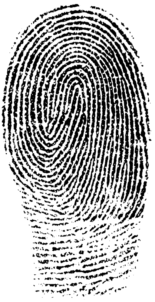
Fingerprinting involves identifying an individual by their unique pattern of arches, loops, and whorls on the ridges of the fingertips. Invisible oils and other biomolecules are laid down on a surface, and crime scene techs visualize those scant traces through a number of methods. Those prints are then compared to a local, national or international database and, hopefully, a match is made and a perpetrator is identified.

But a person’s identity may not be the only thing revealed by his fingerprints, as was recently announced:

  1. Determining the use of illegal drugs: Researchers from the University of Surrey in England have developed a method to test the residue left in a fingerprint for cocaine using mass spectrometry. More importantly, based on the drug metabolites, it can be determined whether the cocaine was ingested, or whether the suspect simply touched it and traces of the drug remained on his fingertips. New portable mass spectrometers are being developed to make this a technique that can be used in the field at actual crime scenes.
  2. Fingerprint Molecular Identification (FMI) technology to identify gender, narcotics and nicotine: North Carolina’s ArroGen Group has developed FMI technology, again using mass spectrometry, to identify gender biomarkers, as well as metabolites of nicotine, heroin, methamphetamine, marijuana, temazepam, ecstasy and even some legal medications. This panel of distinctive chemical substances could lead to suspect identification as well as criminal convictions.
  3. Determining the age of a fingerprint: Researchers at the National Institute of Standards and Technology have developed a method to approximate the age of a fingerprint. This has always been a problem using fingerprints as criminal evidence—a print might prove an individual was in a particular location, or touched a particular object, but was it at the time of a crime, or the week before and therefore possibly insignificant? Scientists have tried to develop a method to date fingerprints based on the breakdown of the biochemical products in the fingerprint, but to no avail. However this method is different and depends on the movement of biomolecules from the ridge to the empty valley sections of the print. Essentially the clearly defined lines in a fresh print will blur and become indistinct with time. How much so will help scientists date an individual print. So far, scientists have been able to distinguish between a day and a week old, a week and a month old, and a month and four months old. This is still a proof-of-concept method, but researchers are working to fine tune the technique, which could be incredibly useful in criminal investigations.
  4. Determining race of an individual: We’ve previously discussed how to determine race from a victim’s skull, but researchers from North Carolina State University recently announced a technique to determine race from the minutiae of the fingerprint. In a nutshell, there are three levels of examination for a fingerprint. The first level is the one most people are familiar with—those ridge formations called arches, loops, and whorls shown in a standard ink print. The second level is the minutiae—the deviations of those arches, loops, and whorls—where a ridge ends, when it splits in two at a bifurcation, or where the ridge makes a U-turn in a loop or whorl. In comparing those from African-American and European-American backgrounds, researchers found significant differences at the minutiae level, enough to be able to determine from which group the individual came. The study only involved 243 subjects, so these are very preliminary results, but so far the data appears promising.

It is early days so far for many of these techniques, but, with additional study, hopefully they will develop into full-fledged tools for investigators, providing them with more information and hopefully leading to more definitive suspect and victim identifications.

Photo credit: Wikimedia Commons

The Word on the Street Toronto 2015

This past Sunday, I once again attended The Word on the Street Toronto, the free annual festival celebrating the best of Canadian writing and reading. This year, for the first time, WOTS took place at Harbourfront Centre, right on the shore of Lake Ontario. A beautiful venue, a stunning day and books for any and all tastes - it was guaranteed to be a good time!

The ride in the on the train proved to be quite rowdy as it was full of Blue Jays fans headed to the Rogers Centre for the final game in a weekend series against the Tampa Bay Rays. The Jays are currently in first place in the American League East Division and have their sights set on the League title and then the World Series. It's been 22 years since they made the playoffs (when they last won the World Series) and Toronto has been in a sports drought for a very long time (the Leafs last won the Stanley Cup in 1967) so fans are extremely excited to have an successful home town team. Did I mention the trip in was rowdy?

A beautiful day at Harbourfront Centre, more reminiscent of summer than fall:

The festival at a glance - so much bookish awesomeness all squeezed together in one park:

All aspects of publishing and reading were represented at WOTS. There were publishers and presses, both large and small in attendance.

The Harlequin tent was always hopping:

HarperCollins:

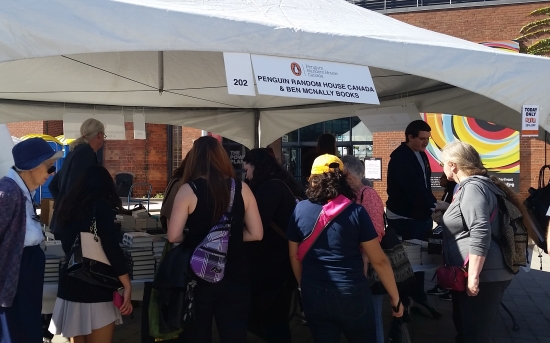
Penguin Random House with Ben McNally Books:

Dundurn Press, the largest Canadian owned publisher:

Montreal comic publisher, Drawn and Quarterly:

And many more...

Both the Toronto Public Library and the TPL workers had booths:

Several bookstores were in attendance, including Mabel's Fables, a well-known local children's bookstore:

Many writing associations and organizations attended, showcasing their authors and their books, as well as their services. I helped man the Crime Writers of Canada booth for part of the day.

The Writer's Community of York Region:

The Writer's Community of Durham Region:

 The Ontario Writers' Conference and The Writers' Union of Canada:

Toronto Sisters In Crime and the Toronto Romance Writers:

Canadian Authors Association and the Science Ficton Writers:

There were even some other tents that made for a nice eclectic mix.

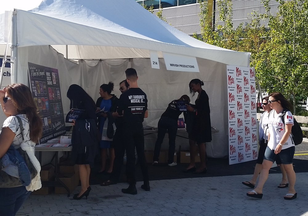
The theater crowd with Mirvish Productions, the biggest theater production company in town:

The Royal Ontario Museum was on hand with several centurions, promoting their terrific Pompeii exhibit:

And Amnesty International had a tent showcasing some of the many ways they work to expose and prevent human rights abuses:

There were a number of tents with presentations ranging from live readings to discussions of current events to awards:

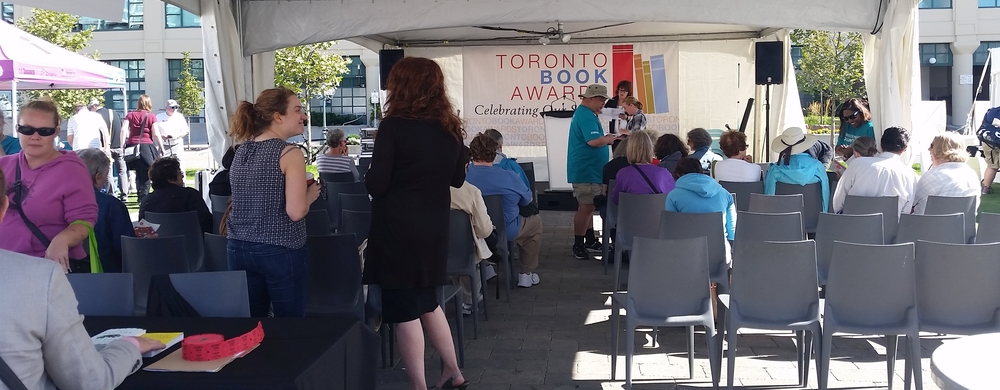
We had a very good crowd pretty much the whole time, but I took advantage of a quick break at one point to grab a picture of the Crime Writers of Canada booth. Lovely authors Sharon Crawford and Caro Soles were signing with me.


Thanks to the Crime Writers of Canada for organizing our part in WOTS and thanks to the many readers who came out to meet new and favourite authors and purchase their work. We all had a great time and are looking forward to next year!

Medieval Skeleton Discovered After Irish Tree Falls

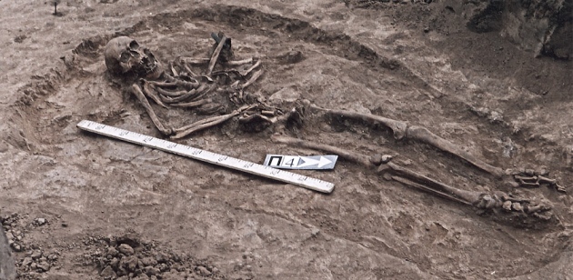
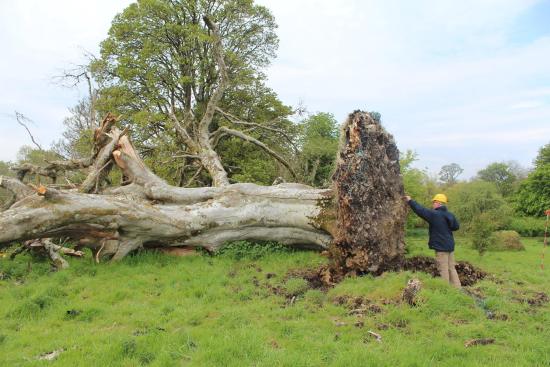
Most weeks this story would be considered firmly based in the past, but compared to our last story concerning possibly three million year old skeletal remains, this archeological find is practically modern. Last week a story appeared on media outlets detailing a 215 year-old beech tree felled by a storm last May in the town of Collooney on the northwest coast of Ireland. Not such a remarkable story (although a shame to lose such an old tree), except for the fact that when the tree uprooted, it revealed a 1,000 year old skeleton beneath. Even stranger, when the tree fell, it ripped the skeleton in half, leaving the lower body still in ground, while the upper body and skull remained wrapped in the roots.

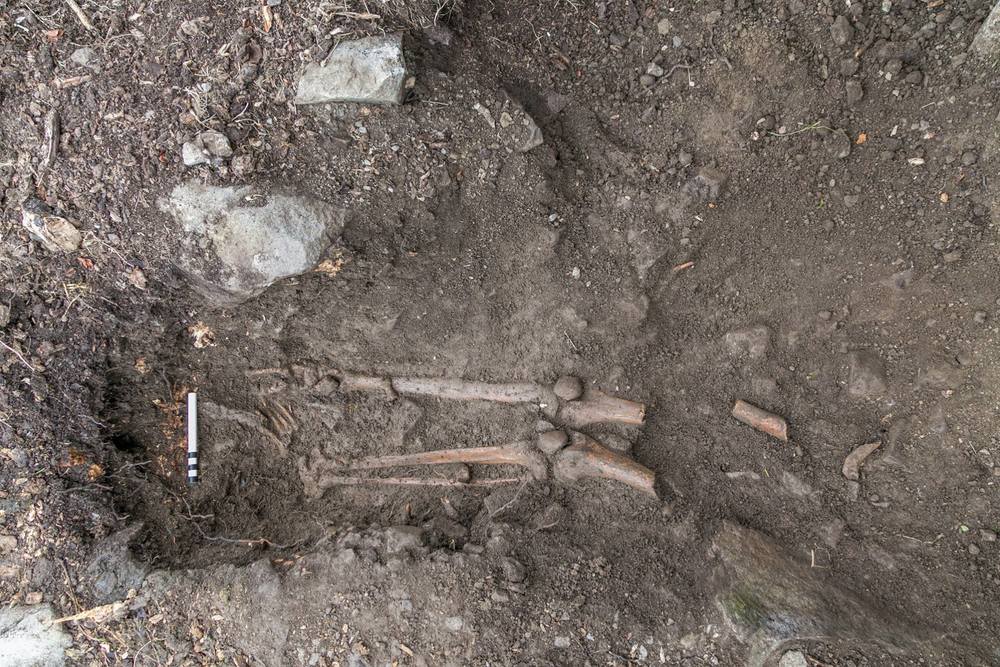
Ireland’s National Monument Service hired the newly formed Slig-Leitrim Archaeological Services to excavate and date the remains as their very first project. Archeologists from Slig-Leitrim determined the remains belonged to a young Gaelic man between the ages of 17 and 20 who died a violent death—sharp force kerf marks were found on the hands and ribs, likely from a sword or knife. While the hand wounds certainly indicate the young man attempted to defend himself, it’s unclear at this time whether he was murdered or died in battle. What is clear from the grave, however, is that it was a formal Christian burial. While there don’t appear to be any other remains in the vicinity, historical records of the area indicate there might have been a church and graveyard in that area long ago.

A human spine tangled in the roots of a felled beech tree.

Radiocarbon (C-14) dating places the bones between 1030 and 1200 A.D., so the young man was buried more than 800 years before the tree sprouted. As the tree grew, the remains of the man’s upper body were ensnared in the roots, so when the tree blew over centuries later, the bones of the upper body were raised into the air, leaving the legs below, still embedded in the ground.

The lower legs and feet still in the ground.

Further analyses of the remains are ongoing, but, once complete, the skeleton will be sent to the National Museum of Ireland, Dublin to be added to their collection.

Photo credit: Thorsten Kahlert

A New Ancestor for Homo sapiens

There’s a lot about human history that modern man doesn’t understand yet. When I was doing research for our story last spring on the oldest known murder, research on Homo heidelbergensis led to somewhat murky conclusions as to its place on the evolutionary timeline. H. heidelbergensis is related to us enough to share the same genus (Homo), but whether we evolved from them or on a completely separate, parallel branch of the ancestral tree is unclear. But last week, a new piece of our ancestral puzzle slid into place when a huge story broke about the discovery of yet another human relative.

Two recreational cavers, Steven Tucker and Rick Hunter, were exploring a cave called the Rising Star, located thirty miles northwest of Johannesburg, South Africa, in an area called the Cradle of Humankind because of the sheer number of fossils of early man discovered there during the early twentieth century. Chances of finding new fossils a century later were minimal, but they were willing to give it a try.

The Rising Star is known for its accessibility only to the most slender and wiry of cavers. It has several passages that narrow to a mere seven and a half inches in width, so exploration of the cave is somewhat restricted. But when these two men passed beyond these significant hurdles and found themselves in the final chamber, they discovered a scattering of ancient bones lying on the sedimentary surface of the cave.

Dr. Lee Burger, a paleoanthropologist working at University of the Witwatersrand in Johannesburg, had asked area cavers to watch for fossils since he lacked the skills for extreme caving. His work involved the mysteries surrounding the evolution of the genus Homo two to three million years ago. Our most distant known relative is Australopithecus afarensis, of which Lucy is the most well-known example. Homo erectus is our nearest relative, but it is within the span between these two species that much of the murkiness, including Homo heidelbergensis, exists. Dr. Berger was convinced that missing pieces to the puzzle were still out there, just still hidden from view. When Tucker and Hunter showed him pictures of the Rising Star cavern, Berger knew that he had to act quickly before any other amateur cavers discovered what he knew to be primitive bones.

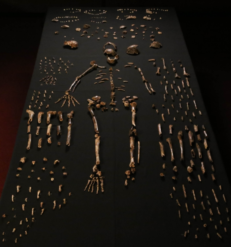
Knowing that Tucker and Hunter didn’t have the skills for an excavation, and that he lacked the skills and physique to enter the cavern himself, Berger put out the call for scientists experienced in both excavation and caving, and with a body form that would allow them entrance into the cavern. He chose six young women from nearly ten times that number of applicants. Over the next four weeks, as Berger and a team of scientists looked on from above with the help of over two miles of data and power cables, the women worked in rotating teams to excavate and remove more than 1,550 bones from fifteen individuals ranging in age from infant to adult.

Dr. Berger called in established scientists from all over the world to help with the skeletal analysis. As an aside, the scientist in me was thrilled to see that he also invited newly minted Ph.D. graduates to participate in what will likely be the find of their lives. The skeletons were divided into ‘workstations’ for each speciality—skulls, teeth, peripheral bones etc. And what they found was remarkable. The specimens were a bizarre combination of humanoid and primitive structures. While the finger bones were curved, indicating tree climbing was a crucial part of their existence, the opposable thumb, palm and wrist bones looked nearly modern. The shoulders and hip bones of the pelvis resembled Australopithecus afarensis’s Lucy, but the bottom of the pelvis and the lower legs and feet resembled modern man. While this was a species with bodies meant for climbing, they also had the long limbs and appropriate muscle attachment points for a bipedal gait.

But, the skulls were significantly different. Four skulls were found—two males and two females. The most notable difference is the size of the braincase—only 51 – 62% of the size of the modern braincase—with an accompanying tiny brain. This alone marks them as non-human since the human brain in all its wonderful complexity is what makes us the species at the top of the food chain, not by sheer strength, where we would not win, but because of our reasoning skills. Dr. Berger dubbed the species Homo naledi as naledi means ‘star’ in Soltho (a local South African language), a reference for the discovery in the Rising Star cave.

The mixed soil composition at the site of the dig has made dating the bones difficult, but researchers have hope that more complex methods may still be used to determine their age. As a result, scientists propose three different niches where H. naledi might have existed:

  1. If the bones prove to be older than three million years, then H. naledi would have co-existed with Australopithecus afarensis’s Lucy, perhaps negating the current theory that Lucy is our oldest relative.
  2. If the bones are between two and three million years old, then H. naledi is likely to be a transitional species between Australopithecus and our own genus Homo.
  3. If the bones are shown to be less than one million years old, then H. naledi may have co-existed with H. heidelbergensis, H. neanderthalensis, and even possibly with the very first Homo sapiens.

Another interesting facet of the discovery of the bones was their location in such a difficult to reach site. The distribution of the bones suggested they were deposited there purposely, and over a long period of time. The lack of animal tooth scoring on the bones indicated that their deaths were not due to animal attack. There was also no trace of ocean sediment to suggest the bodies might have been washed there accidentally. In the end, researchers concluded the bodies were placed there on purpose as part of a primitive funeral ceremony, unexpectedly advanced behaviour for such a species, and possibly the oldest known example of that behaviour in the ancestral human record.

Dr. Lee Berger is a remarkable scientist, more interested in his own research than the celebrity it might bring him. He involved other scientists whose specific knowledge exceeded his own in the interest of the best possible analysis, and he included junior scientists so they learned from the best and could share new out-of-the-box ideas. He waits patiently for the dating results to coalesce instead of jumping to headline-worthy conclusions, and he doesn’t seem interested in overturning current paleoanthropology as we know it. Instead, he pursues science for science’s own sake, and lets the truth of the data lead the way. Among the astonishing data he has presented to us, he may be the most impressive part of this new discovery. As a scientist myself, I take my hat off to you, Dr. Berger. Well done, indeed.

Want more reading? See the original scientific publication here, or the excellent National Geographic article of the discovery here.

Photo credit: Berger et al. in eLIFE

International Literacy Day

Hi all! Sorry to have been away so long, but we've literally just yesterday finished the first draft of LONE WOLF and I've been head down writing like a fiend all summer. We'll be back next week again with our normal blog posts, but this infographic came into my inbox this afternoon, and I thought it would be great to share it. Literacy is such a fundamental skill and the stats outlined certainly show that as a global population we have a long way to go. Take a look...

See you back again next week when we're back and rolling on Skeleton Keys as usual!

Literacy Day

Biosecurity Incidents In Top U.S. Labs—What, Me Worry? Smallpox Edition

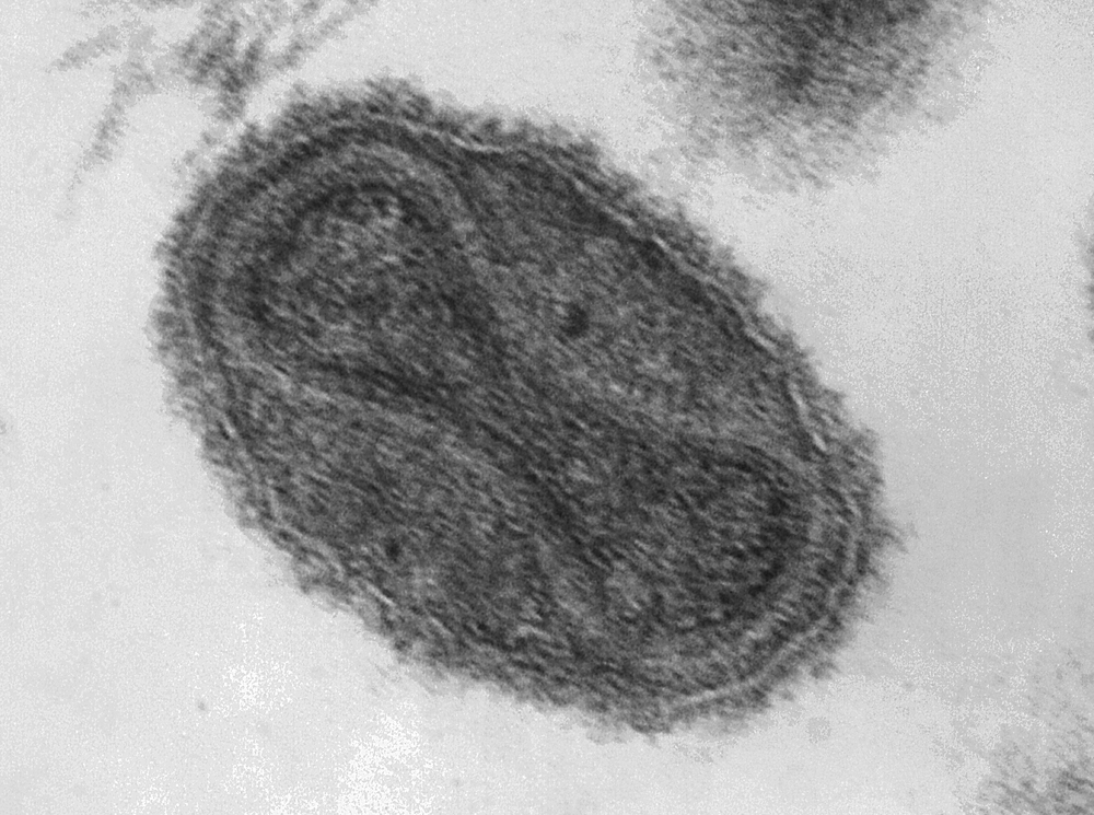
Smallpox is one of the few diseases that modern medical science has managed to eradicate. Believed to have emerged around 10,000 B.C., the disease first appeared in historical documents in the 15th century. The disease is estimated to have killed 300 to 500 million people in the 20th century alone, having a fatality rate of 20–60% in adults and 80% in children.

Smallpox is widely taught in university immunology classes as the world’s first vaccine. Edward Jenner noticed in 1798 that milkmaids who developed cowpox seemed to be immune to smallpox infection. In a stunningly unethical, off-the-cuff experiment, when a milkmaid came to him for treatment, he ran a thread through one of her pustules, coating it with pus. He then inoculated the eight year old son of his gardener by making a small cut and running the pus-coated thread through it. Shortly after, the boy became symptomatic for a mild case of cowpox. Several months later, Jenner took pox scabs from someone with small pox and similarly inoculated the boy a second time. The boy was immune to smallpox and remained healthy. This was the beginning of the vaccine revolution.

In 1967, the WHO mandated the eradication of smallpox, using newer, modern vaccines based on vaccinia virus, a virus related to both smallpox and cowpox. The last known case of smallpox occurred in 1977, and the WHO considered it eradicated in 1979.

This left the world with only laboratory strains of the virus. Following a breach of containment, resulting in the death of a lab worker in 1978, any labs with remaining virus either destroyed them or transferred them to safer labs. Currently, only the U.S. Centers for Disease Control and the Russian State Research Center for Virology and Biotechnology still retain samples. The argument has been made that all remaining virus should be destroyed, but the existing aliquots are retained in case any other stocks arise leading to a dangerous bioweapon in the wrong hands. At this point, decades after the final vaccinations, essentially everyone except military personnel (who continue to be inoculated with the vaccinia vaccine for out of country work) would be susceptible to a fresh onslaught of smallpox. But with only two stocks of the virus in the world, we’d like to believe that we’re safe.

So it was somewhat of a shock in July 2014, when the National Institutes of Health reported that six glass vials of freeze-dried smallpox stock had been found in a long forgotten box in the back of a cold storage room. I remember hearing the news and being stunned for several reasons—glass vials to store a biosafety level IV pathogen (of course, there was no sterile, disposable Nalgene polypropylene cryovials back then, but glass? So incredibly dangerous…), no security, and no inventory so no one even knew they were there. The stock was estimated to have been there since the 1950s, even though the building didn’t open until the 1960s, and from the 1970s on was used by the Food and Drug Administration. A further investigation reveals twelve boxes in total containing smallpox, dengue, influenza, Q fever, and rickettsia, all previously unknown to be stored there, and all with no security precautions (proper security precautions would involve a minimum of two locked doors between the pathogen and the public, detailed inventories, and full biosafety training of all personnel). The FDA immediately mandated a full review of all cold storage spaces to ensure no other pathogens were present.

The glass vials were immediately transferred to the CDC to undergo testing, where it was determined that two of the six vials contained viable virus. Had the tubes broken, the world could have seen a smallpox pandemic the likes of which it hadn’t seen for decades and for which we are entirely unprepared. Luckily for all of us, the vials remained intact. All the vials were destroyed following testing.


Ann and I are going to be taking some time off for summer holidays and to really concentrate on drafting LONE WOLF, book one of the new FBI K-9 Mysteries with Kensington Books. So we look forward to coming back fresh and with a lot of solid writing behind us in September. See you then!

Photo credit: Wikimedia Commons

Biosecurity Incidents In Top U.S. Labs—What, Me Worry? Influenza Edition

In the same report published by the CDC in July 2014 that discussed a laboratory incident concerning anthrax, an incident concerning influenza was also revealed. In August 2014, a full report detailed the cross-contamination of a non-pathogenic H9N2 strain of avian (bird) influenza with the highly pathogenic H1N1 strain (remember the 2009 flu pandemic? That’s the one…) which then sent it out to a U.S. Department of Agriculture lab that had no idea what they were dealing with. Fortunately, the Department of Agriculture lab is also a biosafety level III (BSL III) lab, so the samples were handled under BSL III containment procedures. As it turned out, no workers were infected with the pathogenic strain, a very lucky break as the story could have ended very differently.

As a scientist, the moment I heard this story, I knew exactly what had likely happened and it’s a big no-no in working with cells and viruses. Keep in mind, the error wasn’t identified until six months after the fact, so the worker couldn’t recall the day like it was the previous week, but reported working with then non-pathogenic H9N2 virus, decontaminating the biological cabinet and the working with the pathogenic H1N1 virus. However, of the 1.5 hours required to do all of this, key card access indicated the worker was only present for 51 minutes and that also included time to shower out of the facility and dress in street clothes. Clearly, the full protocol was not carried out. The scientists admitted that they were under pressure at the time to complete work for an upcoming WHO vaccine conference, and, that day in particular, the scientist in question was rushing to get to a lab meeting. Corners were clearly cut.

There are two scenarios that could have happened:

  1. Considering that each infection should take 30 minutes, it is possible the scientist did both infections in the biological cabinet at the same time. To put it plainly, this is NOT done. One of the first rules of tissue culture is that products are kept separate to ensure purity of the product and safety of the current and any future lab worker. I’d like to think this isn’t what happened.
  2. Instead of following protocol and working with the less pathogenic strain first and then the pathogenic strain, the scientist may have mixed up the order, working with the more pathogenic strain first and then not decontaminating afterward before moving onto the second, less virulent strain. Personally, I think this is what happened. Also, as PCR testing of the H9N2 strain doesn’t indicated H1N1 contamination, it’s likely they weren’t used concurrently.

One other concerning incident happened in association with this cross-contamination. When the receiving lab started to use the virus, chickens in the experiment started to unexpectedly die. Upon testing their virus, they determined that their H9N2 was contaminated with the deadly H1N1. When they informed the CDC, the lab team tested their stock of H9N2 to confirm that it was indeed contaminated with H1N1. But they did not report the incident at the time. It wasn’t until a second CDC team found atypical results with their stock of H9N2 virus that the original team reported the incident. At that time, all connected stocks of H9N2 were destroyed. Luckily all work done by the second team was conducted under BSL III containment, so there was no risk to any of the lab personnel.

For a group that is considered by most scientists as the gold standard, this incident combined with the anthrax incident is quite distressing. Scientists did not display good laboratory practices, training, communication skills, and in many cases, common sense. As a scientist myself, especially as one trained in BSL III procedures, many of these errors would simply not be acceptable or expected at our facility. Protocols and training are already in place to avoid this and a detailed incident reporting procedure is in place (and let me assure you, I’ve used it for BSL III incidents twice). After all this, I know I certainly look at the CDC differently, and I’m sure many other scientists do as well.

Next week we’ll be back with our last installment in this series. What happens when a virus that is nearly eradicated pops up in someone’s freezer? Come back next week and we’ll tell you all about it…

Photo credit: Wikimedia Commons

Biosecurity Incidents In Top U.S. Labs—What, Me Worry? Anthrax Edition

Today we’re continuing on with our series on laboratory biosafety and how it’s gone wrong lately in places you would think would be immune to such incidents. But it just goes to show human error can trump every precaution you put in place and that hopefully your people would follow to the letter. Especially when lives are on the line. But not so much, apparently…

In July of 2014, the Centers for Disease Control (CDC) held a press conference to discuss two different incidents that had taken place the previous month. We’re going to showcase one incident this week and one next week.

The first incident involved a CDC lab preparing extracts from anthrax, a biosafety level III (BSL III) pathogen. Their usual protocol involved chemically deactivating the pathogen for 24 hours, but they learned of a new protocol that only required a 10 minute deactivation. Now, this protocol was not for anthrax, but was instead for Brucella, another BSL III pathogen. However, they elected to try this new protocol on anthrax. This method also included a double check—after the pathogen is deactivated, some of it is plated onto agar plates and incubated for 48 hours to ensure there is no growth, and all the pathogen is dead. The CDC first attempted this protocol in June of 2014, sampling the extract to agar plates after 10 minutes, but leaving the rest for the full 24 hours.

However, due to a misunderstanding during a phone conversation and a lack of follow-up with the actual printed protocol, the scientist responsible only left the agar plates for 24 hours post deactivation, at which time, the plates were determined to have no growth. The scientist intended to autoclave the plates and discard them that day, however he could not open the autoclave door, so the plates were returned to the incubator. At this time, the anthrax incubated for 24 hours was distributed as deactivated pathogen to biosafety level II (BSL II) labs. Eight days later, the agar plates were removed from the incubator for disposal and, to their surprise, anthrax growth was observed. Scientists realized the 10 minute procedure was not sufficient to kill anthrax, but were unsure if the 24 hour procedure (from which anthrax had been sent out to BSL II labs and their lower level of containment) was sufficient. At that point, it had to be considered that they had a breach in biosafety containment and all the labs had to be completely decontaminated. Later tests showed that the 24 hour procedure was sufficient to kill most of the anthrax, but not all. As a result, while it was unlikely that infection would occur, it was not impossible.

Upon closer examination, there were a number of issues that led up to this incident:

  1. Use of unapproved techniques—there were several related to filtering of the extract, but this also included observing the plates for sterility at 24 hours instead of the required 48 hours.
  2. Transfer of material not confirmed to be inactive—based on the error made in point 1, this also involved a lack of written protocol of what was required to ensure that pathogens were truly deactivated.
  3. Use of pathogenic strains of anthrax to test out a new protocol when non-pathogenic strains would have revealed the same conclusion with none of the risk. This was an extremely unwise decision.
  4. Inadequate knowledge of peer-reviewed literature by both the laboratory scientist and his supervisor—papers already existed outlining that this method was not sufficient for absolute sterility of infectious anthrax.
  5. Lack of standard operating procedures to document pathogen deactivation.

Fortunately, no staff member ever presented with symptoms of anthrax. But the laboratory in question was closed pending a number of assessments, the establishment of new procedures, and until remedial action was taken with the staff involved. This was not a shining moment for the CDC, the institution we scientists like to consider the gold standard in biosafety. This is the group you call when something goes wrong and this is what their own people are doing?

In the last month, a new anthrax story surfaced concerning an Army lab at the Dugway Proving Ground in Utah. It creates anthrax test kits and sends them out to numerous laboratories for local testing. Contained in the kit is a radiation-killed sample of anthrax to use as a positive control for testing. However, in May, a Maryland biotech company identified that live anthrax was present in the samples. Upon closer inspection, it appears the samples were not sufficiently treated to kill all the spores. The test kits were packaged and sent out, many by regular FedEx delivery, to 69 labs around the United States, but also in Canada, Britain, Australia and South Korea. The CDC has confirmed that all kits shipped between 2004 and 2015 contained live anthrax.

Even more disturbing information surfaced this past week. It appears that from 2007 onwards, the lab was aware that their deactivation process (chemical deactivation at the time, and later irradiation) was not sufficient to kill all the anthrax, but they ignored the issue. At the time, federal regulators at the Office of the Inspector General were aware and recommended further investigation and potential enforcement action, however nothing was ever done. Furthermore, the information was never reported to Congress—the group responsible for oversight of this lab—so they were completely unaware of the situation. No laboratory workers have shown symptoms of anthrax; however 31 workers are receiving antibiotics as a precaution.

Next week, we’ll be returning the CDC as we discuss a serious mishap concerning a highly pathogenic strain of influenza.

Photo credit: Wikimedia Commons

A New Publishing Contract and Format For TWO PARTS BLOODY MURDER

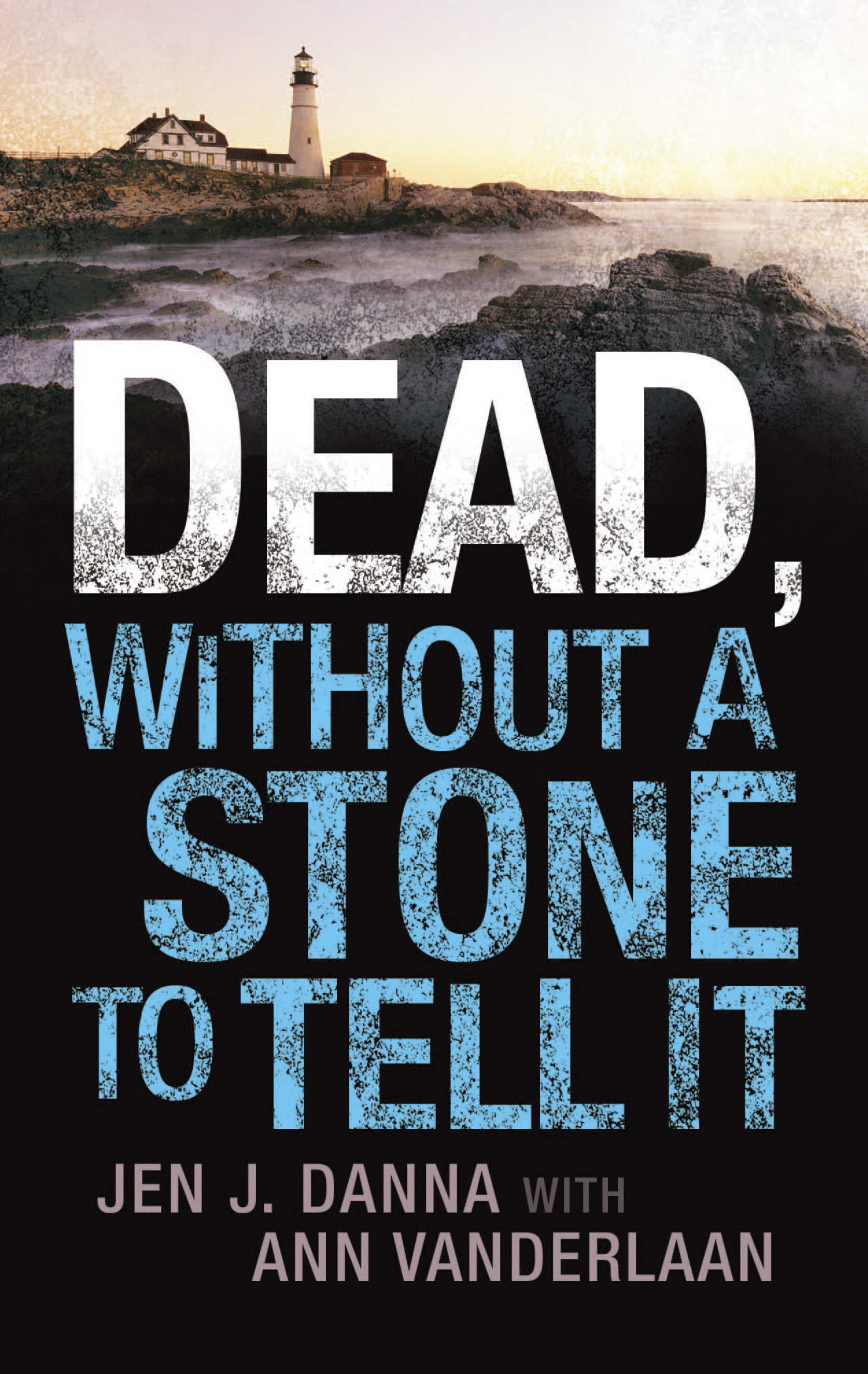
We’re very happy to announce today that TWO PARTS BLOODY MURDER will be joining the ranks of DEAD, WITHOUT A STONE TO TELL IT and A FLAME IN THE WIND OF DEATH as part of Harlequin’s Worldwide Mysteries in mass market paperback format. We’ve been very pleased with our sales through Worldwide and are looking forward to our continuing partnership with them. Currently the projected publication date is spring 2016, but I’ll post the exact date on the website when we know it.

The deal was announced two days ago on Publisher’s Marketplace:

June 21, 2015 - TWO PARTS BLOODY MURDER by Jen J. Danna with Ann Vanderlaan

Jen Danna with Ann Vanderlaan's TWO PARTS BLOODY MURDER, the third novel in the Abbott and Lowell Forensic Mysteries, where a body discovered in a long-forgotten speakeasy proves to be ground zero for a cascade of murders through the decades, to Laura Barth at HQN Worldwide Mystery, for publication in spring 2016, by Nicole Resciniti at The Seymour Agency.

One of the fun aspects of working with Harlequin is that they always do their own back cover blurbs and cover art, so it’s always interesting to see their take on our series. As you can see above, they do an excellent job with series branding, so we can’t wait to see where they take TWO PARTS BLOODY MURDER. So we’ll be back with more on this release in the future as we have more information to share.